European Society of Radiology: Sports imaging is the main theme of IDoR 2019. In most countries, this is not a specialty in itself, but a focus within musculoskeletal radiology. In your country, is there a special focus on sports imaging within radiology training or special courses for interested radiologists?
Hermant Patel: India is rapidly catching up with the western world in the field of medicine and is at par in many urban centres. Musculoskeletal (MSK) radiology is emerging as the top subspecialty of choice among budding radiologists. This is in great part due to an increasing inculcation of sports in the younger generation’s daily routine and wider opportunities available for professional sports practice in the country. This new trend has increased the number of recreational, amateur and professional sports-related injuries that we encounter in our daily practice, and hence the need for expertise in diagnosing and managing these conditions. Sports imaging is emerging as a subspecialty branch of radiology that caters to the MSK imaging needs of athletes of all shapes and sizes, ranging from national cricket, badminton and tennis players, Olympic competitors, serious marathon runners, recreational joggers and people who simply wish to improve their general fitness.
Many elite universities and hospitals in India are coming up with structured fellowship programmes for the subspecialty of sports imaging throughout their MSK training, and outreach programmes are conducted by the Indian Radiological and Imaging Association (IRIA) to update new radiologists with the latest developments in the field.
ESR: Does sports-related imaging take up all, most, or only part of your regular work schedule?
HP: I run a chain of independent private diagnostic centres across the Ahmedabad & Mehsana districts of Gujarat state. Reflecting the growing need for MSK imaging subspecialists, MSK radiology now comprises almost 40–50% of our daily workload, of which about 20–30% are related to sports imaging.
ESR: Based on your experience, which sports produce the most injuries that require medical imaging? Have you seen any changes in this regard during your career? What areas/types of injuries provide the greatest challenge to radiologists?
HP: In our country, cricket is probably the most beloved sport among the young and the old. It is also the most common recreational sport played among the masses, besides tennis or badminton, and the most common cause of sports-related injuries in our daily practice. The recent trend of recreational jogging, ‘gymming’ and marathon running, and the overzealous attempts to attain fit shape in short intervals have also boosted imaging needs.
The greatest challenge to a radiologist in sports imaging is diagnosing field injuries because it puts pressure on us. Imaging findings, besides clinical assessment, strongly influence appropriate management decisions, return to training and competition, and prediction of injury recurrence or risk of serious injury. It is also important to keep in mind that it is difficult to apply a standard imaging finding to all athletes when they vary greatly in muscle range and function, which is required to perform specific activities in different sports. This diversity is reflected in the heterogeneity of muscle injuries. Another challenging arena is diagnosing paediatric trauma.
I have always found that understanding the relevance of imaging findings is easier when accompanied by the knowledge of anatomy, biomechanics and pathological processes involved in injury formation, allowing us to form a rational conclusion.
ESR: Please give a detailed overview of the sports injuries with which you are most familiar and their respective modalities.
HP: Knee menisci and ligamentous injuries are probably the most common injuries I encounter the most, especially amongst cricketers and young football players. Acute shoulder dislocation and rotator cuff tears are quite common in young bowlers and avid golfers. Ankle ligamentous injuries are most frequent amongst badminton and tennis players, besides young skaters and gymnasts. There is also an increase in the traumatic finger injuries that are imaged, possibly due to now available treatment and rehabilitation options. MR imaging is the modality of choice, yielding the maximum diagnostic information in these cases.
With the rising trend of ‘gymming’ and large breed of fitness fanatics, a large number of patients end up with muscle injuries, due to overenthusiastic usage of gym equipment.
Ultrasound and magnetic resonance imaging (MRI) are the most frequently applied imaging modalities in sports medicine.
ESR: What diseases associated with sporting activity can be detected with imaging? Can you provide examples?
HP: Sports imaging can largely be divided into two categories: those that are due to overuse, i.e. repetitive action such as tendinositis, arthritis and stress fractures; and those which are traumatic, such as shoulder dislocation or fractures. With new technologies in ultrasound, MRI and multidetector computed tomography (MDCT), the possibility to document subtle findings has been pushed forward enormously. This has led to the detection of slight stress-related marrow changes, typical patterns of tendinositis in overuse syndromes, muscle trauma or early forms of marrow contusions. Distinct sports-related signs have been described using new insights in the pathophysiology. Imaging is crucial to assess the extent of sports-related muscle injuries and help guide management, which directly affects prognosis. This is especially important when the diagnosis or grade of injury is unclear, when recovery is taking longer than expected, and when interventional or surgical management could be considered.
Along with the detection of early abnormalities, a new field of research is also emerging for the prevention of tendon ruptures, osteoarthritis and fractures.
ESR: Radiologists are part of a team; for sports imaging this likely consists of surgeons, orthopaedists, cardiologists and/or neurologists. How would you define the role of the radiologist within this team, and how would you describe the cooperation between radiologists, surgeons, and other physicians?
HP: In sports-related injuries, the main goal of the sports medicine team is to return the athlete to competition – balanced against the need to prevent the injury from worsening or recurring. Prognosis based on the available clinical and imaging information is crucial. Imaging is crucial to confirm and assess the extent of sports-related injuries and guide further management, which directly affects prognosis.
Radiologists play a pivotal role in sports medicine. Following clinical assessment, many athletes will require some form of imaging to confirm the suspected diagnosis and assess its severity, for example, an x-ray examination to exclude a fracture, MRI scan to diagnose a meniscal or cartilage injury or ultrasound to assess the hamstring muscle injury. Thus, sports imaging is an important link between sports medicine and imaging sciences, stimulating each other within interdisciplinary clinical management.
ESR: The role of the radiologist in determining diagnoses with sports imaging is obvious; how much involvement is there regarding treatment and follow-up?
HP: Sports imaging expertise is not limited to diagnosing these conditions; it is also useful in treating and preventing them. Radiologists increasingly perform treatment and prevention interventions, e.g. pain management interventions, platelet-rich plasma (PRP) injection of a muscle tear, dry needling or cortisol injection in tenosynovitis, cortisol injections into an inflamed joint or harvested tendon cells (tenocytes) into a damaged Achilles tendon. These procedures are performed under ultrasound or fluoroscopy guidance by a trained interventional radiologist.
ESR: Radiology is effective in identifying and treating sports-related injuries and diseases, but can it also be used to prevent them? Can the information provided by medical imaging be used to enhance the performance of athletes?
HP: The primary goal of athletes is to minimise their ‘on the bench’ time and return to training and competition as soon as possible. High re-injury rates remain a major problem following acute injuries. Re-injuries are often more severe than the initial injury and are associated with longer absence from sports.
Although clinical examination remains the core of any patient assessment, radiology plays an increasingly important role in the initial assessment and follow-up of these injuries. Follow-up imaging is particularly helpful in the longitudinal assessment of grade 2 injuries exhibiting partial muscle fibre disruption whenever clinical symptoms persist after proper management and rehabilitation, as well as in cases of a clinical suspicion of re-injury.
Many sports medicine physicians truly appreciate high-quality imaging to help guide athlete rehabilitation, although the clinical evaluation largely guides the final return-to-play decision.
ESR: Many elite sports centres use cutting-edge medical imaging equipment and attract talented radiologists to operate it. Are you involved with such centres? How can the knowledge acquired in this setting be used to benefit all patients?
HP: High-end ultrasound machines with newer probes that allow high-resolution imaging for dynamic muscle and tendon evaluation and nerve assessment, as well as MR machines with higher-strength magnets and dedicated coils for small joint examination, are routinely used in our diagnostic centre. We have often had the opportunity to image elite national and international players, as we are part of the sports imaging partners for the Indian cricket premier league whenever it is played in our city. Cutting edge technology and properly trained radiologists in sports imaging allow us to cater to these athletes in the best possible manner.
ESR: The demand for imaging studies has been rising steadily over the past decades, placing strain on healthcare budgets. Has the demand also increased in sports medicine? What can be done to better justify imaging requests and make the most of available resources?
HP: Clinical assessment remains the core of any patient assessment and should be duly supported by imaging analysis wherever applicable. However, imaging alone should never be used as a substitute for clinical assessment. It is important to understand the relevance of the imaging findings in a given clinical scenario.
ESR: Athletes are more prone to injuries that require medical imaging. How much greater is their risk of developing diseases related to frequent exposure to radiation and what can be done to limit the negative impacts from overexposure?
HP: The most common modalities used are ultrasonography and MR imaging, which do not use ionising radiation, whereas x-ray related radiation exposure would probably never cross the occupational radiation hazard limits. Care can be undertaken while performing computed tomography (CT) studies and limiting the radiation exposure to the area of suspicion and using lead safety aprons to cover the rest of the body. Unfortunately, awareness of radiologists and sports medicine consultants is low regarding the importance of radiation safety and judicious usage can limit the risk of harmful effects.
ESR: Do you actively practise sports yourself and if yes, does this help you in your daily work as MSK radiologist?
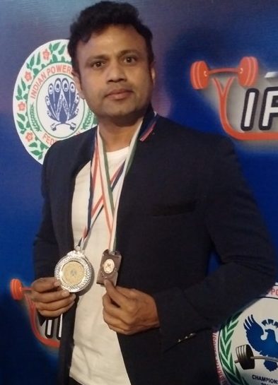
HP: I am part of the Indian team for powerlifting and have won many medals in this discipline. I represented India and won the Silver Medal at the Open International Power Lifting Championship in Jamshedpur in January 2016. In 2016 I developed some nagging pain in my right front shoulder. I underwent MRI and found out some marrow oedema surrounding acromioclavicular joint. This was due to heavy bench-pressing. I started with regular warm and band exercise before my lifting sessions and could get rid of the pain permanently. This is a common thing to happen because back muscle develops very fast whereas pectorals may take some years to catch up to maintain equilibrium. Similarly, due to heavy deadlifting and squat, one may develop lower backache, due to wrong form/technique or overuse.
I strongly recommend proper technique, form and warmups to all my radiologists and physician friends practising sports. I also advise listening to your body and taking conscious rest for a while if your body shouts for any pain due to overuse.
European Society of Radiology: Sports imaging also applies to sports-related injuries of the brain. In case you are familiar with this, please also answer the following questions:
ESR: Which sports have the highest risk of inducing brain injuries?
HP: Although sports injuries rarely contribute to fatalities, the leading cause of death from sports-related injuries is traumatic brain injury. The risk is highest amongst contact sports like football, boxing and wrestling. Recently there have been incidents of young cricketers suffering from a fatal head injury due to cricket ball impact.
ESR: What imaging modalities do you use with traumatic brain injury specifically in athletes?
HP: Sports-related head injuries, including acute subdural haematoma (ASDH), traumatic cerebrovascular disease, cerebral concussion, and chronic traumatic encephalopathy (CTE). In the medical setting, head CT or MRI should be performed to rule out organic lesions. Diagnostic imaging is highly recommended to exclude the possibility of brain injuries such as ASDH and cerebral haemorrhage, especially when the individual has a persistent headache.
ESR: What can be learned from sports-related injuries that can be applied to a broader use, for example those sustained through automobile or other accidents that cause traumatic brain injury?
HP: Traumatic brain injuries by automobiles and sports occur by almost the same mechanism in terms of sudden rotational acceleration/deceleration or high impact and have a similar appearance in diagnostic imaging, though with much more intensity for automobile accidents. Such accidents are also more associated with polytrauma and spinal injuries.
ESR: How have advances in brain imaging allowed you to predict patient outcomes more accurately?
HP: Advanced neuroimaging of concussive injury is a growing field within sports-related concussion research. Routine neuroimaging studies, including cranial MRI and CT, are typical in the majority of concussed patients, leading many to call concussion a functional rather than structural injury. However, there has been research using more advanced MRI protocols, particularly diffusion tensor imaging (DTI), which can demonstrate white matter tract disruptions and help in prognosis.
 Dr. Hermant Patel is Professor of Radiology and Managing Director at Gujarat Imaging Centre, Post-Graduate Institute of Radiology & Imaging in Ahmedabad. He is the president of the Indian Radiological and Imaging Association (IRIA).
Prof. Patel is also part of the Indian team for powerlifting and has won many medals in this discipline, including the Silver Medal at the Open International Power Lifting Championship in Jamshedpur, India, in January 2016.
Prof. Patel is an invited speaker for teaching programmes in the USA, Europe, China, Korea and SAARC countries. He has delivered more than 350 lectures as a guest speaker at various conferences in India and has given more than 40 presentations at the RSNA and 20 presentations at the ECR. Prof. Patel is a primary author in many international peer-reviewed journal publications, including international journals like Radiographics and Neurographics. He has also published extensively in national journals.
Dr. Hermant Patel is Professor of Radiology and Managing Director at Gujarat Imaging Centre, Post-Graduate Institute of Radiology & Imaging in Ahmedabad. He is the president of the Indian Radiological and Imaging Association (IRIA).
Prof. Patel is also part of the Indian team for powerlifting and has won many medals in this discipline, including the Silver Medal at the Open International Power Lifting Championship in Jamshedpur, India, in January 2016.
Prof. Patel is an invited speaker for teaching programmes in the USA, Europe, China, Korea and SAARC countries. He has delivered more than 350 lectures as a guest speaker at various conferences in India and has given more than 40 presentations at the RSNA and 20 presentations at the ECR. Prof. Patel is a primary author in many international peer-reviewed journal publications, including international journals like Radiographics and Neurographics. He has also published extensively in national journals.