European Society of Radiology: Sports imaging is the main theme of IDoR 2019. In most countries, this is not a specialty in itself, but a focus within musculoskeletal radiology. In your country, is there a special focus on sports imaging within radiology training or special courses for interested radiologists?
Can Çevikol: In Turkey, sports imaging is not a subspecialty itself. However, it is within the scope of musculoskeletal (MSK) radiology as a part of radiology training. The Turkish Society of Radiology (TSR) and Turkish Society of Magnetic Resonance frequently organise MSK courses for interested radiologists where sports imaging is usually included in the programme.
ESR: Please describe your regular working environment (hospital, private practice). Does sports-related imaging take up all, most, or only part of your regular work schedule?
CÇ: I work in the Akdeniz University Hospital in Antalya, Turkey. It is a reference hospital with a 1,050 bed capacity. Around 12 million tourists visit the Akdeniz Region every year. Especially in the early season and mid-season, many sports clubs come to Antalya for training and therefore our hospital serves thousands of patients from all over Turkey and other neighbouring countries. I work in the MRI department at the hospital and my primary area of interest is MSK radiology. Sports imaging is a part of my daily, routine practice.
ESR: Based on your experience, which sports produce the most injuries that require medical imaging? Have you seen any changes in this regard during your career? What areas/types of injuries provide the greatest challenge to radiologists?
CÇ: Regarding my experience, injuries requiring medical imaging occur mostly in contact sports such as football, basketball and wrestling. During winter, injuries from skiing and snowboarding tend to increase. In addition, oil wrestling competitions, which is a traditional Turkish sport, are commonly held in southern Turkey which cause many spine, knee and shoulder injuries.
Throughout the span of my 25-year career, our society has become more fitness oriented than ever and a large part of the general population is involved in recreational sport activities. Nowadays, we see more injuries related to recreational sports activities. I think this underlines the importance of sports medicine and medical imaging in our daily practice. In my opinion, the greatest challenge for general radiologists is mostly related to the imaging of hand and fingers due to their anatomic complexity and the difficulty of obtaining standard high-quality images.
ESR: Please give a detailed overview of the sports injuries with which you are most familiar and their respective modalities.
CÇ: I mostly see ankle, knee, hip and shoulder pathologies related to sports in my daily practice. Furthermore, I am mostly familiar with ligamentous, muscle, tendinous and bony injuries of these anatomical regions. In general, conventional radiography should always be the first diagnostic modality to depict bone or joint abnormalities. For evaluation of fractures and fracture healing, CT is the preferred modality. For the diagnosis of muscle and tendon problems, ultrasound (US) is considered the best imaging modality but, it is user dependent and experience is very important for correct diagnosis. US and MRI constitute most of my daily, routine practice. Achilles tendon pathologies, plantar fasciitis, tendon and ligament problems such as rotator cuff disease, tenosynovitis of any superficial tendon, synovial cysts, inguinal hernias, avulsion injuries and bursitis are well-known indications for use of ultrasound. MRI of the knee is the most frequently requested study for knee derangement in our department. MRI is the preferred imaging modality for deep-lying tendons and intra-articular ligaments, providing excellent sensitivity and specificity. It can also identify the other source of pain-like degeneration and muscle problems. Therefore, osteochondral lesions, labral abnormalities, ligamentous and tendinous injuries, avascular necrosis, stress fractures, bone marrow abnormalities, muscle strains and meniscus problems are evaluated with MRI.
ESR: What diseases associated with sporting activity can be detected with imaging? Can you provide examples?
CÇ: In addition to findings related to acute injury, chronic overuse injuries such as myositis ossificans, osteitis pubis and sport hernia can be detected with imaging. Additionally, sports-related brain injuries can be detected. Since they are not recommended as a first-line screening tool, cardiac MRI, coronary calcium scoring, and computed tomography coronary angiography have also been promoted as potentially valuable screening tools for competitive athletes.
Regular and prolonged exercise can cause increase in the thickness of the left ventricular wall that can overlap with hypertrophic cardiomyopathy (HCM) findings. Differentiating physiological from pathological hypertrophy is important, since HCM is the most common cause of exercise-related sudden cardiac death in young individuals.
ESR: Radiologists are part of a team; for sports imaging this likely consists of surgeons, orthopaedists, cardiologists and/or neurologists. How would you define the role of the radiologist within this team and how would you describe the cooperation between radiologists, surgeons, and other physicians?
CÇ: The role of the radiologist is to make correct and fast diagnosis, which is important in deciding treatment options and especially preventing unnecessary surgery. In elite athletes, the time of recovery and return to sport should be decided by the radiologists and orthopaedic surgeons.
ESR: The role of the radiologist in determining diagnoses with sports imaging is obvious; how much involvement is there regarding treatment and follow-up?
CÇ: Regarding treatment, image-guided, especially ultrasound-guided interventional procedures such as drainage of collections and cysts may be used. For sclerosis of neovascularity in painful chronic tendinosis, IR has also been described for pain reduction. Ultrasound is especially useful for the follow-up of muscle strains. MRI can be used for the follow-up of stress injuries, bone marrow oedema, muscle strains, microfractures, osteochondral lesions, as well for the postsurgical evaluation.
ESR: Radiology is effective in identifying and treating sports-related injuries and diseases, but can it also be used to pre-empt them? Can the information provided by medical imaging be used to enhance the performance of athletes?
CÇ: Recent advances in imaging technologies may provide relevant data about those subjects. Examples include diffusion tensor imaging (DTI) which shows muscle architecture and structure, monitoring subtle changes in skeletal muscle related to age, atrophy or disease. T2-mapping, spectroscopy, blood-oxygenation-level-dependent (BOLD) imaging, and molecular imaging have the potential for better understanding muscle biomechanics, muscle energetics, and joint function.
ESR: Many elite sports centres use cutting-edge medical imaging equipment and attract talented radiologists to operate it. Are you involved with such centres? How can the knowledge acquired in this setting be used to benefit all patients?
CÇ: I have not worked with an elite sports centre before. There is one private elite sports centre in Antalya. Professional Turkish and European sport teams visit this centre in winter due to the warm weather of the city, but this centre doesn’t have its own medical imaging equipment. As far as I know, they collaborate with private medical centres for occasional emergencies.
ESR: The demand for imaging studies has been rising steadily over the past decades, placing strain on healthcare budgets. Has the demand also increased in sports medicine? What can be done to better justify imaging requests and make the most of available resources?
CÇ: It is true that the demand for imaging studies has been increasing, including studies in sports medicine. Imaging does not replace the need for good clinical evaluation. Therefore, to better justify the imaging studies, all patients should be evaluated by experienced sports physicians or orthopaedists. Another important issue regarding making the most of available resources is reducing unnecessary examinations.
ESR: Athletes are more prone to injuries that require medical imaging. How much greater is their risk of developing diseases related to frequent exposure to radiation and what can be done to limit the negative impacts from overexposure?
CÇ: It is well known that ionising radiation is harmful and there is no safe lower threshold of radiation due to stochastic effects. Therefore, care must be taken about the radiation dose to the patient when radiography or a CT examination is performed. Children require higher levels of consideration since they are at greater risk from radiation than adults. Therefore, when appropriate, the alternative use of safer non-ionising techniques such as US and MRI should be preferred. Especially in children and adolescents, low dose techniques for radiography and CT should be considered.
ESR: Do you actively practise sports yourself and if yes, does this help you in your daily work as MSK radiologist?
CÇ: In the past, I have done many sports activities such as tennis, swimming, and basketball as a recreational activity. During that period, I experienced many muscle, bone and tendon injuries. This situation helps me to better understand the injured athlete, and I can put myself in his or her situation. It also improves my understanding of the relevance of minor injuries in professional athletes which may be irrelevant in non-athletic patients. Therefore, I give additional caution to the detection of minor findings.
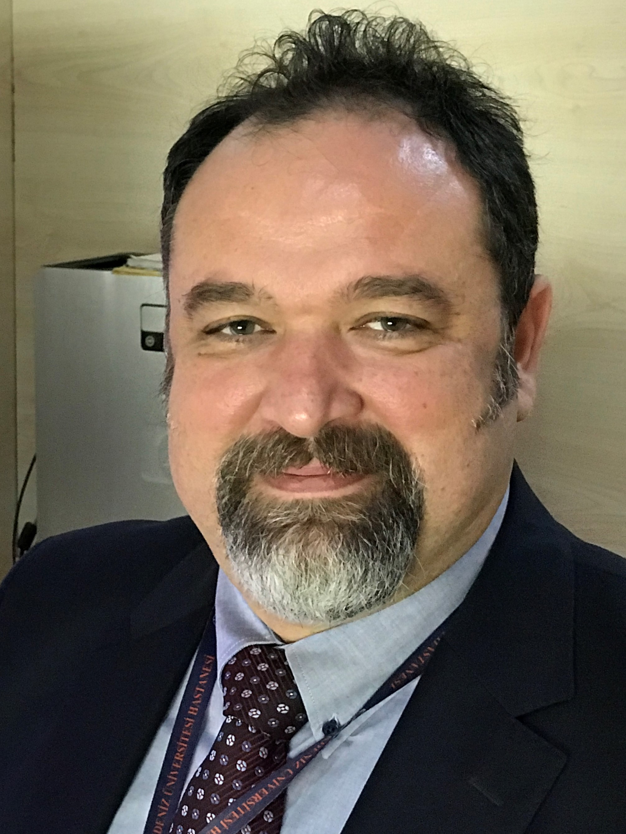 Prof. Can Çevikol is a musculoskeletal radiologist at Akdeniz University in Antalya, Turkey. He graduated from Hacettepe University Faculty of Medicine (1991) in Ankara and completed his radiology training in Akdeniz University Faculty of Medicine (1997) in Antalya.
He is member of the European Society of Radiology (ESR), European Society of Musculoskeletal Radiology (ESSR), Balkan Society of Radiology (BSR) and Turkish Society of Magnetic Resonance (TSMR). He is the author of 50 scientific papers published in peer-reviewed journals and more than 100 presentations at international and Turkish Congresses. His scientific papers have 274 citations. Dr. Çevikol is currently Vice President of the Turkish Society of Radiology.
Prof. Can Çevikol is a musculoskeletal radiologist at Akdeniz University in Antalya, Turkey. He graduated from Hacettepe University Faculty of Medicine (1991) in Ankara and completed his radiology training in Akdeniz University Faculty of Medicine (1997) in Antalya.
He is member of the European Society of Radiology (ESR), European Society of Musculoskeletal Radiology (ESSR), Balkan Society of Radiology (BSR) and Turkish Society of Magnetic Resonance (TSMR). He is the author of 50 scientific papers published in peer-reviewed journals and more than 100 presentations at international and Turkish Congresses. His scientific papers have 274 citations. Dr. Çevikol is currently Vice President of the Turkish Society of Radiology.