European Society of Radiology: Could you please give a detailed overview of when and for which diseases you use cardiac imaging? Which modalities are usually used for what?
Jonathon A. Leipsic: Cardiac CT has rapidly evolved from a test that was used to complement other testing done for the evaluation of patients with chest pain and suspected coronary artery disease to the first line test for this purpose. It has been shown to be the most accurate and prognostically informative test and now, through large-scale randomised trials, it has been shown that CCTA guided decision making helps improve clinical outcomes with a significant reduction in downstream heart attacks. Combined with new techniques, integrating advanced computational modelling CTA can also provide the haemodynamic significance of coronary narrowings fractional flow reserve (FFR) CT.
We also use cardiac CT to guide minimally invasive transcatheter valvular interventions to improve outcomes.
Cardiac MRI also has growing evidence supporting its role as an essential tool for the workup and management of heart failure patients, patients with congenital heart disease, and to help guide revascularisation strategies.
ESR: What is the role of the radiologist within the ‘heart team’? How would you describe the cooperation between radiologists, cardiologists, and other physicians?
JAL: The heart team is an important multidisciplinary team that has had a renaissance with the advent of transcatheter aortic valve replacement (TAVR). The radiologist and other cardiac imagers play an integral role in these important discussions and in guiding decision making.
ESR: Radiographers/radiological technologists are also part of the team. When and how do you interact with them?
JAL: Cardiac imaging is dependent on excellent image quality which is equally dependent on thoughtful, committed and well-trained technologists. We work closely with the technologists in serving our patients and driving improvements in image quality.
ESR: Please describe your regular working environment (hospital, private practice). Does cardiac imaging take up all, most, or only part of your regular work schedule? How many radiologists are dedicated to cardiac imaging in your team?
JAL: We have a team of five radiologists and one cardiologist focusing on cardiac CT and MRI. Our team is deeply embedded in our Heart Centre and we play an integral role in diagnosing and guiding treatment decision making.
ESR: Do you have direct contact with patients and if yes, what is the nature of that contact?
JAL: Our coronary CT slate is typically 15–18 per day. We greet the patients and take a clinical history to understand the nature of their symptoms to help make more sense of their coronary artery findings and to help with planning downstream treatments.
ESR: If you had the means: what would you change in education, training and daily practice in cardiac imaging?
JAL: I can continue to work hard academically to advance practice and work with others to continue to define the future role of cardiac imaging for the betterment of the patients we serve.
ESR: What are the most recent advances in cardiac imaging and what significance do they have for improving healthcare?
JAL: The recent introduction of FFR CT has very much changed how we practice. For the first time with a resting coronary CT, we can obtain both anatomy and physiology allowing us to not only diagnose coronary disease but also determine which patients need to be revascularised.
ESR: In what ways has the specialty changed since you started? And where do you see the most important developments in the next ten years?
JAL: Change is a constant in medicine and the field of cardiac imaging is no exception.
ESR: Is artificial intelligence already having an impact on cardiac imaging and how do you see that developing in the future?
JAL: We are seeing and we have published about the use of large registry data and deep learning to define novel predictors of a downstream heart attack. We are also seeing the maturation of tools like FFR CT using AI and integrating invasive physiology to further optimise and improve the test. The future is bright and opportunities endless.
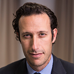
 Professor Jonathon A. Leipsic, MD, FRCPC FSCCT is Professor of Radiology and Cardiology at the University of British Columbia in Vancouver, Canada. He is the Chairman of the Department of Radiology for Providence Health Care and the Vice Chairman of Research for the UBC Department of Radiology. Dr. Leipsic is also a Canada Research Chair in Advanced Cardiopulmonary Imaging. He recently has taken on the role of Medical Imaging Regional Department Head of Vancouver Coastal Health.
Professor Leipsic has over 415 peer-reviewed manuscripts in press or in print, over 300 scientific abstracts, and is editor of 2 textbooks. He speaks internationally on a number of cardiopulmonary imaging topics with over 150 invited lectures in the last four years and is Past President of the Society of Cardiovascular CT, the largest international society focused on cardiovascular CT.