European Society of Radiology: Could you please give a detailed overview of when and for which diseases you use cardiac imaging?
Alejandro Zuluaga: The most frequent heart diseases in our service are coronary ischaemic disease, dilated cardiomyopathy, asymmetric hypertrophic cardiomyopathy, acute and chronic myocarditis, non-ischaemic cardiomyopathies (including amyloidosis, sarcoidosis, Anderson-Fabry’s disease, non-compact left ventricle and eosinophilic cardiomyopathy), congenital cardiac malformations, cardiac masses and pulmonary hypertension.
The main clinical indications in our cardiovascular imaging section are:
- Suspicion of coronary ischaemic disease in asymptomatic patients
- Suspicion of coronary ischaemic disease in symptomatic patients
- Assessment of ventricular systolic function in cases where echocardiography is not conclusive
- Differentiation between ischaemic and non-ischaemic cardiomyopathy
- Viability assessment
- To help define the aetiology of non-ischaemic cardiomyopathy according to the pattern of late gadolinium enhancement (LGE) or the parametric values of T1, T2, T2* in cardiac magnetic resonance (CMR)
- To establish the presence, extent and location of left ventricular myocardial fibrosis so as to define patient prognosis and help guide electrophysiology procedures when indicated (resynchronisation therapy/ablation therapy/implantation of implantable cardio defibrillator)
- Establish the diagnosis of causes of MINOCA (myocardial infarction with non-obstructive coronary arteries) such as acute myocarditis, stress-induced cardiomyopathy (Takotsubo cardiomyopathy) or acute myocardial infarction of embolic origin or with spontaneous recanalisation
ESR: Which modalities are usually used for what?
AZ: I work in a private ambulatory imaging diagnostic centre (CEDIMED) with an academic agreement with two universities (Universidad Pontificia Bolivariana UPB and Universidad CES) in Medellin. In our cardiovascular imaging section, we have two magnetic resonance imaging (MRI) scanners – one 3T and one 1.5T – and a 64-multidetector computed tomography (MDCT) scanner. In the following section, we will describe the main clinical indications for performing cardiovascular studies with our MDCT and MRI equipment.
Cardiac MR (CMR):
In patients with suspected non-ischaemic cardiomyopathy, with asymmetrical hypertrophic cardiomyopathy or with dilated cardiomyopathy, we use contrast-enhanced CMR, including anatomical sequences, cine sequences to assess ventricular function; 2D and 3D late gadolinium enhancement (LGE) sequences to evaluate late enhancement pattern to define the aetiology of cardiomyopathy and to define the extent and precise location of fibrosis; T1, T2, T2* mapping and extracellular volume percentage to help diagnose infiltrative processes such as amyloidosis, Anderson-Fabry’s disease and iron overload cardiomyopathy. In patients with iron overload, studies are performed on a 1.5T MRI scanner.
In symptomatic patients with suspected coronary ischaemic disease with intermediate pre-test risk (15–85%), we use contrasted enhanced cardiac MRI, including anatomical sequences; cine sequences; perfusion sequence with stress (adenosine) to assess perfusion disorders during stress that present resolution at rest; 2D and 3D LGE sequences to evaluate areas of old infarction and viability criteria.
In a patient with a clinical MINOCA, we use contrast-enhanced CMR, including anatomical sequences; cine sequences to assess ventricular function and regional motility disorders of the left ventricle wall (either from myocardial infarction or Takotsubo cardiomyopathy); T2 sequences to assess myocardial oedema; 2D and 3D LGE sequences to evaluate late enhancement pattern for either myocarditis or myocardial infarction; T1, T2 and percentage extracellular volume mapping to aid in the diagnosis of acute myocarditis or other non-ischaemic cardiomyopathies.
In patients with ventricular arrhythmia in which we want to define and characterise the presence of left or right ventricular fibrosis in order to guide ablation therapy or to define the patient’s prognosis and establish the need to implant a cardio-defibrillator, we use contrast-enhanced cardiac MRI, which includes anatomical sequences; cine sequences; 2D and 3D LGE sequences to define the presence of fibrosis by either ischaemic or non-ischaemic pathology, defining fibrosis extent (percentage), precise location (endocardial or epicardial) and determining the presence and location of border zone channels (slow conduction channels).
Cardiac MDCT:
In asymptomatic patients with suspected coronary ischaemic heart disease with intermediate risk of coronary heart disease due to clinical rates/scores or with a significant family history of coronary heart disease, we use cardiac CT for calcium scoring.
In symptomatic patients with suspected coronary ischaemic heart disease with an intermediate pre-test probability of coronary heart disease, we use coronary computed tomography angiography (CCTA).
In the evaluation of the patient prior to a transcatheter aortic valve implant (TAVI), we use contrast cardiac CT with electrocardiographic synchronisation to evaluate the characteristics of the aortic valve, aortic root, coronary arteries, and left ventricular outflow tract. In addition, we perform a CTA of the abdominal and thoracic aorta without electrocardiographic synchronisation to evaluate the procedure approach.
In the evaluation of the patient with atrial fibrillation before or after ablation therapy, we perform mapping and evaluation of the left atrium and pulmonary veins, we use contrast-enhanced cardiac CT with electrocardiographic synchronisation.
ESR: What is the role of the radiologist within the ‘heart team’? How would you describe the cooperation between radiologists, cardiologists, and other physicians?
AZ: We are two cardiovascular imaging trained radiologists (Natalia Aldana Sepúlveda, MD and myself) who perform the interpretation of cardiac MRI and MDCT studies at our institution. The role of the radiologist in our cardiovascular imaging section includes evaluation of the medical orders for CMR and CCTA studies, to define if the indication of the study is appropriate. We also coordinate the scheduling of studies, define patient preparation for the exams, and evaluate patients’ medical history and previous exams. Radiologists are present during CCTA and CMR studies to define the protocol to be performed in each patient according to the clinical indication of the examination. In addition, we perform post-processing of images, and interpret the studies and communicate the results to the treating physicians when required.
ESR: Radiographers/radiological technologists are also part of the team. When and how do you interact with them?
AZ: Besides us radiologists, our team includes four MRI technologists and one MDCT technologist. The cardiac team technologists have received training in cardiovascular imaging at our own institution.
ESR: Does cardiac imaging take up all, most, or only part of your regular work schedule? How many radiologists are dedicated to cardiac imaging in your team?
AZ: Our institution is an outpatient diagnostic imaging centre with 33 radiologists who are part of the different subspecialties of diagnostic imaging. We the radiologists in charge of cardiovascular imaging devote approximately 25% of our regular working hours to this field.
ESR: Do you have direct contact with patients and if yes, what is the nature of that contact?
AZ: Yes, radiologists in charge of cardiac imaging studies are always present during the exams and we have the opportunity to talk to the patients, to explain how the studies are performed, answer their questions and, when necessary, to tell them about the results.
ESR: If you had the means: what would you change in education, training and daily practice in cardiac imaging?
AZ: I consider the concept of an integral approach in non-invasive cardiac imaging to be fundamental, so that the physician who is specialised in this area has sufficient knowledge and training in the different non-invasive diagnostic modalities (MDCT, CMR, SPECT, PET and echocardiography) and plays a more active and effective role in the multidisciplinary team responsible for treating patients with cardiovascular disease.
Following this concept, I believe the objectives should focus on giving radiology residents greater exposure to the basic concepts of cardiac physiology and anatomy and general training in all non-invasive diagnostic modalities such as cardiac MDCT, MRI, echocardiography, SPECT and PET. In addition, cardiac imaging subspecialty programmes should be created where fellows receive in-depth training in all these non-invasive cardiac diagnostic modalities so that they can perform without difficulty in any practice setting, both in the field of cardiology and radiology.
ESR: What are the most recent advances in cardiac imaging and what significance do they have for improving healthcare?
AZ: In CMR, I would highlight the following advances:
- The value of detecting and characterising myocardial fibrosis with late gadolinium enhancement sequences, to establish the prognosis of patients independent of the type of cardiomyopathy, helping to identify patients requiring an implantable cardio defibrillator, to guide ventricular ablation therapy and to guide resynchronisation therapy. In our service, we have two years of experience in the characterisation of myocardial fibrosis using the 3D late gadolinium enhancement sequence with an isotropic high spatial resolution of 1.4 x 1.4 x 1.4 mm obtained on a 3T MR scanner and performing 3D post-processing with special software (ADAS-VT, Galgo Barcelona), which allows precise definition of fibrosis extension, its endo or epicardial location, and the presence of border zone channels. These slow conduction channels have been shown to promote the development of re-entry circuits and are a frequent site of origin of malignant ventricular arrhythmias, therefore becoming the target for ablation of the left ventricle.
- Recent clinical validation demonstrating the current value of cardiac MR myocardial stress perfusion testing, with results similar to or better than the conventional stress echocardiography and stress myocardial perfusion testing with SPECT and PET used so far.
- Recent advances in T1 mapping and extracellular volume quantification techniques in the characterisation of infiltrative cardiomyopathies such as amyloidosis and Anderson-Fabry’s disease
- The usefulness of T2* mapping in the evaluation of patients with iron overload cardiomyopathy
As far as cardiac MDCT is concerned, I would highlight the following advances in recent years:
- A significant decrease in radiation doses, mainly due to different iterative reconstruction techniques
- The possibility to add functional information to cardiac MDCT studies, either through CCTA-derived fractional flow reserve (FFRCT) or through stress perfusion cardiac MDCT studies, increasing the specificity and positive predictive value of CCTA and providing additional essential information to guide the management of patients with suspected ischaemic heart disease. The important limitations of these techniques are their currently poor accessibility, cost factor and the need for broader clinical validation in general non-academic practice.
- Important technological advances such as an increase in the number of detectors that improve coverage with a lower rotation time of the gantry; dual-source CT scanners that improve the temporal resolution of the studies; and dual-energy CT scanners that improve the contrast between different tissues
ESR: In what ways has the specialty changed since you started? And where do you see the most important developments in the next ten years?
AZ: First I would highlight the clinical validation of tissue characterisation techniques either with LGE sequences or with T1, T2 and T2* mapping that have exponentially increased the indications for CMR, including evaluation of ischaemic and non-ischaemic cardiomyopathies, myocardial infiltrative pathologies, electrophysiological indications and assessment of patients with acute pathologies such as MINOCA.
I have also been able to appreciate the evolution of the concept of absolute contraindication of cardiac MR in patients with non-conditional implantable cardiac devices (pacemakers and implantable cardio defibrillators) for MRI (legacy devices). According to recent studies that have been performed in specialised centres, it would seem safe to perform cardiac MRI in 1.5T MRI scanners in a selected group of patients with non-conditional MRI devices, according to the specific characteristics of each device, with strict monitoring of patients and following a safety protocol including electrophysiologists and all the personnel in charge of performing the MRI study.
On the other hand, the evolution of the security concept of Gadolinium-based contrast agents (GBCA) has also been evident. Whereas in the past these contrast media were considered ‘very safe’ and were often given in double doses, clinical evidence since 2006 has demonstrated the risk of nephrogenic systemic fibrosis (NSF) in patients with renal failure receiving administration of Gadolinium-based contrast media. Recently it has also been possible to establish the risk of developing gadolinium deposits after the administration of repeated doses of GBCA in the absence of renal failure, initially reported in the central nervous system (basal ganglia and dentate nucleus). Implications and clinical consequences have not been established at the moment, but they have alerted international regulatory agencies and led to the suspension of marketing licenses of some of these contrast media.
I believe the main advances we will experience in the next ten years in non-invasive cardiovascular imaging could be, in MRI: faster image acquisition, for example with compressed sensing techniques; new fast and robust techniques for volumetric (3D) myocardial perfusion assessment; T1 mapping, phase contrast and late enhancement; development of new, safer MRI contrast media for patients; and development of non-breath-hold and non-ECG-gated CMR techniques.
In MDCT, I believe advances will be made in clinical validation and standardisation of current FFR techniques derived from CT; in new techniques to value the FFR derived from CT by the different commercial companies of unrestricted use; and in new myocardial perfusion techniques for MDCT using less radiation.
ESR: Is artificial intelligence already having an impact on cardiac imaging and how do you see that developing in the future?
AZ: An example of the impact of artificial intelligence (AI) on cardiac imaging can be seen in our daily practice in the latest version of the software we use in post-processing and interpretation of our cardiac MR studies. For the development of this new version of the software, they used deep learning (in particular Convolutional Neural Networks, CNN) in nearly 5,000 cardiac MRI studies to train ventricular segmentation in ventricular function analysis.
I definitely believe that in the near future we will see the influence of AI in all phases of our work in cardiac imaging, including image acquisition, post-processing and study interpretation.
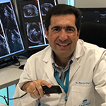
 Dr. Alejandro Zuluaga, MD, FSCMR, FSCCT is a radiologist in the diagnostic medical centre CEDIMED and associate professor of radiology at the Universidad Pontificia Bolivariana (UPB) and Universidad CES in Medellin, Colombia. He is also director of the radiology residence programme in the UPB.
Dr. Zuluaga received his medical degree and radiology degree from the Universidad CES. He completed a subspecialisation in trauma and emergency radiology at the Jackson Memorial Hospital – Miami University in Miami, Florida, USA.
Dr. Zuluaga worked for three years as an associate professor of radiology at the body imaging section of Louisiana State University (LSU) in New Orleans, USA. In 2006, he completed a fellowship in cardiovascular imaging at Stanford University in Palo Alto, California, USA.
Since 1995, Dr. Zuluaga has been an active researcher in trauma and emergency radiology, with a special focus in helical CT and MDCT of cervical trauma, traumatic vascular injuries, ureteral stones, acute abdomen and acute aortic syndromes. Since 2006, he has focused his academic activity on cardiovascular imaging, both CT and MRI. During the last five years, he has been dedicated to research on the usefulness of cardiac CT and cardiac MRI in patients with ventricular and atrial arrhythmias.
Dr. Zuluaga is a recognised speaker and has been invited to present 68 lectures in national and international congresses. He is the author of a book on CT protocols, 20 peer-reviewed articles, 21 book chapters and 45 scientific exhibits and oral presentations.
He was a member of the scientific committee of the Asociación Colombiana de Radiología (ACR) for ten years and sat on the organising committee of two congresses of the Colegio Interamericano de Radiología (CIR) and ten annual meetings of the ACR. He is director of the CIR department of cardiac imaging.