European Society of Radiology: Sports imaging is the main theme of IDoR 2019. In most countries, this is not a specialty in itself, but a focus within musculoskeletal radiology. In your country, is there a special focus on sports imaging within radiology training or special courses for interested radiologists?
Gonzalo Del Carpio-Bellido: I am afraid there is no special focus on sports imaging in Peru or most Latin American countries, with the exception of Argentina, where football goes hand-in-hand with radiology. What a small group of radiologists are trying to do is focus on musculoskeletal issues, but those who are interested in that exciting field mostly need to go outside their countries for training or experience.
ESR: Please describe your regular working environment (hospital, private practice). Does sports-related imaging take up all, most, or only part of your regular work schedule?
GCB: I work in the main private centre in the city of Lima, with five different satellite centres. During my shift, I am assigned to the team of radiologists who are responsible for supervision and interpretation of all general musculoskeletal imaging studies. We also do procedures like arthro-MRI and arthro-CT. A lot of these examinations are requested for injured athletes. We also communicate on a regular basis with their surgeons or sports medicine specialists to comment on treatment and get feedback on surgery findings and outcomes. Sports-related imaging takes up most of my regular work schedule.
ESR: Based on your experience, which sports produce the most injuries that require medical imaging? Have you seen any changes in this regard during your career? What areas/types of injuries provide the greatest challenge to radiologists?
GCB: Almost a third of all musculoskeletal injuries are sports-related, and by far, caused by football practice. In Peru, we now also practise volleyball extensively, but during my career, football-related injuries were the most common. Stress fractures are often a diagnostic challenge since they are not really visible on imaging other than magnetic resonance imaging (MRI).
ESR: Please give a detailed overview of the sports injuries with which you are most familiar and their respective modalities.
GCB: Glenoid labrum lesions are frequent in overhead activities. We have a long and proud tradition of volleyball ‘matadoras’ (female killers) in that field, and the modality of choice is always computed tomography (CT) or MRI arthrogram. The glenoid labrum is also subject to a wide range of normal variants that can mimic labral tears, and the correct technique and knowledge of these variants are central to avoid misdiagnosis. A torn meniscus is visualised every day and most often is not a diagnostic challenge at all. However, from time to time, we do have controversy between a not-so-clear tear versus mucoid degeneration. Multidetector CT arthrogram is the tool we use in those cases. The multi-slice data and sub-millimetre slice thickness give no space for doubt. Imaging of the postoperative knee has also become more common because more arthroscopic repair procedures – instead of the old “take it all out and hope for the best” – are being performed. Specific findings of a re-torn meniscus can only be achieved by arthrogram.
ESR: What diseases associated with sporting activity can be detected with imaging? Can you provide examples?
GCB: The goal of sports medicine is to let the athlete get back to competition and prevent the injury from worsening. The most common injury is muscle strain, mainly in the lower limbs. Although its evaluation by MRI has been extensively described, ultrasound gives us certainly distinct advantages; it is fast and relatively inexpensive, to mention a few. The post-injury assessment is important too. MRI can show findings of chronic muscle injury or complications such as muscle hernias and myositis ossificans.
ESR: Radiologists are part of a team; for sports imaging this likely consists of surgeons, orthopaedists, cardiologists and/or neurologists. How would you define the role of the radiologist within this team, and how would you describe the cooperation between radiologists, surgeons, and other physicians?
GCB: The role of the radiologist within the team is to provide the necessary information so that the surgeon can operate, or so that the clinician can begin timely treatment. If this information is not adequate, the best surgical or therapeutic decisions cannot be made, and the patient’s recovery will be affected. In turn, we benefit from the information obtained from the attending physician about the result of the operation, or the success or failure of a particular treatment – in short, how close to reality was the diagnosis proposed by the radiologist. It is impossible to do a good job being isolated from the rest of the specialties.
ESR: The role of the radiologist in determining diagnoses with sports imaging is obvious; how much involvement is there regarding treatment and follow-up?
GCB: The evolution of sports injuries goes hand in hand with the comparison of images before, during and after treatment, which can be surgery, physical therapy or even just a good period of rest. Tracking the patient through image will not only give an idea of the evolution of their disease, but also when they can resume their competitive activity.
ESR: Radiology is effective in identifying and treating sports-related injuries and diseases, but can it also be used to prevent them? Can the information provided by medical imaging be used to enhance the performance of athletes?
GCB: Sports radiology not only focuses on the diagnosis of injuries, but also on the detection of conditions that can trigger injuries when a certain structure is subjected to an exceptionally increased load or effort. A good example would be the medical examination that precedes big transfer negotiations in professional football. These can vary from club to club, but typically involve cardiac screenings, blood tests and MRI scans. Hips, thighs, knees and legs MRI are useful to assess pre-existent intra and extra-articular disease. A dedicated imaging protocol is needed to get a good overview. The implications of misdiagnosis for the athlete and their coach and team may be catastrophic.
ESR: Many elite sports centres use cutting-edge medical imaging equipment and attract talented radiologists to operate it. Are you involved with such centres? How can the knowledge acquired in this setting be used to benefit all patients?
GCB: I am not familiar with such elite centres. At the moment, radiology applied to sports is a branch that is not very developed in my country, but with the arrival of new equipment and techniques, I see great evolution in the area in the next five or ten years.
ESR: The demand for imaging studies has been rising steadily over the past decades, placing strain on healthcare budgets. Has the demand also increased in sports medicine? What can be done to better justify imaging requests and make the most of available resources?
GCB: This is a complex problem since not only do expenses increase (more orthopaedic surgeons are ordering MRIs and arthrographies), but insurers request very high premiums or simply refuse to insure high-performance athletes. This situation can be improved by optimising imaging expenditures, avoiding unnecessary examinations or using cheaper studies that provide basic information, for example, in the follow-up of a superficial muscle tear. Ultrasound has shown its enormous utility in these specific cases.
ESR: Athletes are more prone to injuries that require medical imaging. How much greater is their risk of developing diseases related to frequent exposure to radiation, and what can be done to limit the negative impacts from overexposure?
GCB: Radiation means x-rays and CT scans, which are mostly avoided when MRI and ultrasound are available. But even when absolutely needed, exposure to radiation can be substantially decreased using lower doses and limiting studies to the area of interest. Non-radiology specialists need to be educated in order to reduce especially CT scans – many are being done unnecessarily – and to decrease the time to perform studies that involve fluoroscopy.
ESR: Do you actively practise sports yourself and if yes, does this help you in your daily work as MSK radiologist?
GCB: I practise swimming and basketball on a regular basis. This helps me particularly to understand the patient who wishes not only to recover as quickly as possible, but to return to the activity that he or she enjoys with the same enthusiasm and intensity as before the injury. Knowing the magnitude of the possible injuries also helps me not to overexert myself in sports, or not to perform activities for which I have not been properly trained. We have an inside joke here at our centre when looking at a particular damaged joint – knee, mostly: “Man was not made to play football”. This isn’t meant to discourage the thousands of people who enjoy this awesome sport, but it serves as a reminder that the body must be prepared for excess.
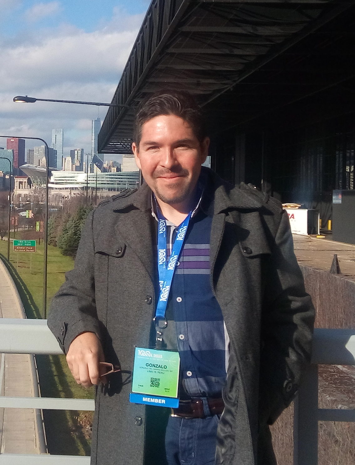 Dr. Gonzalo Del Carpio-Bellido is a radiologist currently working at the Resocentro imaging centre in Lima, Peru. He has a special interest in musculoskeletal imaging and diagnostic procedures. He also serves as second-editor and consultant of the Peruvian Radiology Magazine. During his residency at the Cayetano Heredia Hospital, Dr. Del Carpio-Bellido was known as a pioneer of local online medical blogging. His blog (tiocayetano.blogspot.com), though discontinued in 2009, was one of the first attempts to spread basic radiology knowledge and still offers a nice collection of case studies.