European Society of Radiology: Could you please give a detailed overview of when and for which diseases you use cardiac imaging? Which modalities are usually used for what?
Alexandros Kallifatidis: Cardiac CT (CCT) and cardiac MRI (CMR) have been established as sophisticated advanced imaging techniques and nowadays they play a key role in a wide spectrum of cardiac diseases. Cardiac CT is used mainly in order to rule out or to detect coronary artery disease (CAD) by offering diagnostic and prognostic information, to image bypass grafts and coronary stents but also and in other cardiac pathology such as in patients with congenital heart disease. Cardiac MRI is a very complete and advanced imaging modality playing a leading role in CAD by detecting ischaemia (stress MRI) and revealing myocardial viability as well as in cardiomyopathies and myocarditis with the main advantage of tissue characterisation (late gadolinium enhancement, T1/T2/T2* mapping, oedema sequences etc.). Cardiac MRI plays an additional role in valve disease and other pathologies like tumours, pericardial disease etc. Its role in congenital heart disease is also very important.
ESR: What is the role of the radiologist within the ‘heart team’? How would you describe the cooperation between radiologists, cardiologists, and other physicians?
AK: Radiologists play a fundamental role within the cardiac imaging team. A well-trained and experienced cardiac radiologist offers a sophisticated and holistic diagnostic approach to heart disease. It is important for this approach to take place in the context of teamwork, within the ‘heart team’, together with well-trained cardiologists and in cooperation with physicists and biomedical engineers. A multidisciplinary procedure works better and leads to more effective diagnostic results for the patients and of course for their therapy.
ESR: Radiographers/radiological technologists are also part of the team. When and how do you interact with them?
AK: Well trained and experienced radiographers and technologists are undoubtedly part of the ‘heart team’. Cardiac CT and cardiac MRI are dynamic procedures, personalised for each patient so it is important that everything is set up properly and in a fine collaboration (selected protocols, imaging parameters, radiation dose …).
ESR: Please describe your regular working environment (hospital, private practice). Does cardiac imaging take up all, most, or only part of your regular work schedule? How many radiologists are dedicated to cardiac imaging in your team?
AK: I work in St. Luke’s Hospital, a private hospital in the city of Thessaloniki, one of the biggest in Greece and in South-East Europe. We are two experienced cardiac radiologists in CCT and CMR and one experienced cardiologist in CMR. Cardiac imaging takes up the most of my regular work schedule (both cardiac CT & cardiac MRI). In our department, we perform many of the latest imaging techniques in cardiac radiology, such as CMR stress-perfusion, CMR mapping techniques, CMR strain etc. in daily clinical practice. In addition, we are in collaboration with well-known Greek and European radiology and cardiology departments for research purposes.
ESR: Do you have direct contact with patients and if yes, what is the nature of that contact?
AK: It is really important for clinicians (as we are, clinical radiologists) to develop a relationship of trust with their patients. For me, this relationship is an integral part of my job and of course it helps to have direct contact with patients when discussing the imaging findings and the correlation with the clinical status and the therapy perspective.
ESR: If you had the means: what would you change in education, training and daily practice in cardiac imaging?
AK: Cardiac radiologists should combine deep imaging technique knowledge (physics, acquiring images, protocols, technical parameters etc.) together with deep clinical background and critical thinking. These are the fields that we should emphasise.
ESR: What are the most recent advances in cardiac imaging and what significance do they have for improving healthcare?
AK: Stress tests, mapping techniques, myocardial strain and plaque morphology characterisation are some of the most important advances in cardiac CT and cardiac MRI. Their diagnostic and prognostic significance are very high, offering an impressive healthcare improvement.
ESR: In what ways has the specialty changed since you started? And where do you see the most important developments in the next ten years?
AK: With the rapid technical improvement in the last two decades, and the high quality of the new generation scanners, the progress in the diagnostic quality is spectacular. The coming years are very promising with new applications, new sequences and impressive results in the field of myocardial diffusion, 4D flow, CMR whole heart imaging, hybrid imaging, myocardial spectroscopy, myocardial oxygenation etc. expected.
ESR: Is artificial intelligence already having an impact on cardiac imaging and how do you see that developing in the future?
AK: There is a big discussion and conflicting opinions about the impact of artificial intelligence in general.
Of course, there are a lot of advantages and I believe that AI will play a key role in the future. On the other hand, it is important to realise that the human being is irreplaceable. Medicine is more than technology … It is about the human touch. After all, the answer to every question should be – the human being.
ESR: Please feel free to add any information and thoughts on this topic you would like to share.
AK: Cardiac radiology … the future is here!!!
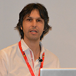
 Dr. Alexandros Kallifatidis is a radiologist with a subspecialty in cardiovascular imaging and a leading specialist in cardiac imaging (cardiac CT, cardiac MRI) in Greece. He is a board-certified cardiovascular imaging radiologist (European Board of Cardiovascular Radiology – EBCR) and an active subcommittee member of the Education & EBCR Committee of the European Society of Cardiovascular Radiology (ESCR).
After finishing his residency in Thessaloniki he was trained in cardiac imaging in Heidelberg, Germany (Cardiology Department, University Hospital Medizinische Klinik with Professor Katus and Professor Korosoglou), as well as in Marseille, France (Radiology Department, University Hospital La Timone with Professor Bartoli and Professor Jacquier).
Since 2007, Dr. Kallifatidis has been working in St. Luke’s Hospital in Thessaloniki, Greece. Under his supervision the Diagnostic Cardiac Section became one of the leading and best-known units in Greece, performing many of the latest imaging techniques in cardiac radiology, such as CMR stress-perfusion, CMR mapping techniques, CMR strain etc. in daily clinical practice. In addition, research collaborations with well-known Greek and European radiology and cardiology departments are performed here.
Dr. Kallifatidis is an active speaker in Greece and Europe and he organises scientific courses in Greece, with real experts and pioneers in the field of cardiovascular imaging as guest lecturers.
Since 2015 he has been collaborating with the Radiology Department of University Hospital CHU-Hôpital Nord in St. Etienne, France under Professor Croisille.
Dr. Kallifatidis is an Executive Committee Member of the Radiological Society of Northern Greece and an Educational Committee Member of the Hellenic Radiological Society.
He is serving as a member of the Programme Planning Committee for ECR (Cardiac Section).