European Society of Radiology: Could you please give a detailed overview of when and for which diseases you use cardiac imaging?
Hajime Sakuma: At Mie University Hospital cardiac imaging is used in all types of cardiac diseases, including suspected coronary artery disease (CAD) in patients with chest pain, dyspnoea, etc.
It is also used in patients after previous myocardial infarction (MI), to determine the indication of revascularisation and in patients with heart failure (HF).
Other indications of cardiac imaging are:
- Assessment of myocardial viability and of the presence and extent of myocardial ischaemia
- Differentiation between ischaemic cardiomyopathy (CAD) and dilated cardiomyopathy (DCM)
- Diagnosis and risk stratification of DCM
- Diagnosis and risk stratification of hypertrophic cardiomyopathy (HCM)
- Diagnosis and risk stratification of myocardial diseases
Cardiac imaging is also key in the following pathologies:
- Cardiac sarcoidosis, cardiac amyloidosis, Fabry’s disease, Takotsubo cardiomyopathy
- Myocarditis
- Pericarditis
- Cardiac tumours and thrombus
- Valvular heart diseases
- Congenital heart disease
- Coronary aneurysms in Kawasaki disease
- Anomalous coronary artery
ESR: Which modalities are usually used for what?
HS: I would first like to emphasise the following unique points in cardiac imaging at Mie University Hospital. Firstly, we routinely perform comprehensive cardiac magnetic resonance (CMR) including whole heart coronary MRA, cine MRI, stress and rest perfusion MRI, T1 mapping, LGE MRI and coronary MRA.
Secondly, we also do stress cardiac CT studies with a radiation dose of <10mSv, calcium scoring, stress perfusion CT, coronary CTA and delayed enhanced CT.
We do ordinary coronary CTA for suspected CAD in patients with chest pain and dyspnoea (among other symptoms), and to rule out CAD with low pre-test probability.
In patients with intermediate to high pre-test probability, we do stress cardiac CT including stress CT perfusion (for ischaemia) and delayed enhanced CT (infarction).
In younger patients and in patients with heart failure, we use comprehensive CMR including coronary MRA.
In patients with dialysis, we do stress single photon emission computed tomography (SPECT). To determine the indication of revascularisation in patients after previous MI, we use stress cardiac CT, including stress CT perfusion (ischaemia) and delayed enhanced CT (infarction).
Generally, for heart failure, the first choice modality is CMR including cine, stress perfusion MRI, T1 mapping, LGE, coronary MRA.
CMR including cine, stress perfusion MRI, T1 mapping, LGE will be used to image DCM HCM and cardiac amyloidosis.
For cardiac sarcoidosis, we prefer CMR including cine, T2W MRI, T1 mapping and LGE / PET/CT for disease activity.
For Fabry’s disease, the first modality of choice is CMR including cine, T1 mapping and LGE, and for Takotsubo cardiomyopathy, we will use CMR including cine, T2W MRI, T1 mapping, stress perfusion, LGE and coronary MRA.
Myocarditis is also best imaged using CMR including cine, T2W MRI, T1 mapping, early enhanced MRI and LGE.
For pericarditis, we can use CT (pericardiac calcification) and CMR including cine and LGE, while cardiac tumours and thrombus are best imaged with CT and CMR including cine, T2W MRI, perfusion MRI and LGE.
Valvular heart diseases and congenital heart disease require using CT and/or CMR.
For coronary aneurysms in Kawasaki disease and anomalous coronary artery, we prefer ultra-low-dose CT and/or coronary MRA.
ESR: What is the role of the radiologist within the ‘heart team’? How would you describe the cooperation between radiologists, cardiologists, and other physicians?
HS: Radiologists play a major role in the heart team and there is an excellent relationship between radiologists and cardiologists. Radiologists perform both CMR and CT studies, including pharmacological stress. Radiologists also write all CMR and cardiac CT studies reports. Good communication between radiologists and cardiologists, who work together on a daily basis, is vital.
ESR: Radiographers/radiological technologists are also part of the team. When and how do you interact with them?
HS: Although the study protocols for both CMR and CT in our hospital are quite complex and operator dependent, technologists and radiologists also have a good relationship, which is based on mutual trust. Fortunately, there are several excellent radiological technologists who have a research mind. When starting new CMR or CT imaging protocols, we radiologists discuss with the technologists quite intensively and evaluate the images together. However, once the imaging protocols are stabilised, we leave actual image acquisition to the technologists.
ESR: Please describe your regular working environment (hospital, private practice). Does cardiac imaging take up all, most, or only part of your regular work schedule? How many radiologists are dedicated to cardiac imaging in your team?
HS: I am working in an academic hospital. As a professor and chairman, as well as deputy director of the university hospital and chairman of the ethical review committee in our institution, the time I have for cardiac imaging is limited. There are eight radiologists in our diagnostic cardiac radiology team, including myself, and we are quite active in clinical research in cardiac imaging. However, all of us are also involved in general radiology reading, such as thoracic and abdominal CT, etc.
ESR: Do you have direct contact with patients and if yes, what is the nature of that contact?
HS: Currently I have no direct contact with patients.
ESR: If you had the means: what would you change in education, training and daily practice in cardiac imaging?
HS: Education and training are very important to foster younger radiologists who will forge the future of cardiac imaging. I was trained in cardiac radiology by performing cardiac catheterisation, coronary angiography, echocardiography, nuclear cardiology and phosphorous cardiac magnetic resonance spectroscopy, which gave me sufficient experience in functional, physiological and metabolic assessment of the heart. Therefore, I frequently suggested young radiologists, who are interested in becoming cardiac radiologists, to work in a cardiology department, to acquire experience in cardiac catheterisation and echocardiography.
However, as the training requirements for board-certified radiologists are getting harder, spending half a year or one year in a cardiology department to broaden one’s perspective and experience is now difficult. I believe such flexibility is good for young radiologists in training, who will play a central role in cardiac imaging for patients.
ESR: What are the most recent advances in cardiac imaging and what significance do they have for improving healthcare?
HS: The most exciting advance is 5D whole-heart self-navigated golden angle MRI. The major limitations of CMR are complexity and operator dependency of CMR data acquisition, and low acquisition efficiency by cardiac and respiratory gating. By utilising compressed sensing for speed and artificial intelligence for automated optimised image reconstruction, 5D (or 6D and 7D) whole-heart self-navigated golden angle MRI will allow us to obtain cine MRI, whole heart coronary MRA (and T1 mapping, etc.) with just a single click of the ‘start acquisition’ button on the MR console. CMR is currently underutilised. This new approach will greatly expand the use of CMR, reduce the radiation dose by substituting x-ray and CT exams, and improve patient care.
ESR: In what ways has the specialty changed since you started? And where do you see the most important developments in the next ten years?
HS: 30 years ago I started cine MRI and P-31 MR spectroscopy of human myocardium. Coronary MRA, my area of expertise in clinical research, also has a history of about 27 years. For the past 30 years, there have been numerous technical developments with so many variations of pulse sequences for CMR. In Japan, there are 52 MR systems per 1,000,000 population, which is the highest in OECD countries. However, the number of CMR studies in Japan is quite limited, being less than one-tenth of coronary CTA. The diversity of pulse sequences in CMR and the operator dependency to utilise them are the major factors restricting the use of CMR in patient care. I believe that convergence and simplicity, instead of divergence and complexity, will be the most important points for the next ten years. From that perspective, the approach such as 5D, 6D or 7D whole-heart self-navigated golden angle MRI is getting more and more important.
ESR: Is artificial intelligence already having an impact on cardiac imaging and how do you see that developing in the future?
HS: Artificial intelligence has not currently actually impacted my practice in cardiac imaging. However, the potential uses of artificial intelligence will increase dramatically. Manual tracing of the endocardial and epicardial border of the myocardium is currently required for the accurate quantification of LV volumes and function. Within a couple of years, this will be replaced by deep learning.
The use of artificial intelligence is essential for image reconstruction as well. For example, artificial intelligence will be a key technique to reconstruct 3D images at systole or diastole, at expiration or inspiration, or to reconstruct excellent coronary MR images from whole-heart self-navigated golden angle MRI dataset.
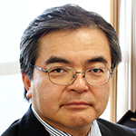
 Professor Hajime Sakuma is professor and chairman of the department of radiology, and deputy director of Mie University Hospital in Tsu, Japan. He graduated from Mie University in 1985. From 1991 to 1996 at the University of California San Francisco, he was engaged in a number of CMR research activities, such as cardiac spectroscopy, k-space segmented fast cine MRI, myocardial perfusion MRI, coronary MRA and coronary flow measurements. He continued and expanded CMR research after moving back to Mie, with major fields of research in MR coronary imaging and myocardial perfusion quantification.
Professor Sakuma is a fellow of the International Society for Magnetic Resonance in Medicine (ISMRM), a fellow of the American Heart Association (AHA), and an honorary member of the European Society of Cardiovascular Radiology (ESCR). He is also President of the Asian Society of Cardiovascular Imaging (ASCI).