European Society of Radiology: Sports imaging is the main theme of IDoR 2019. In most countries, this is not a specialty in itself, but a focus within musculoskeletal radiology. In your country, is there a special focus on sports imaging within radiology training or special courses for interested radiologists?
Juan Cosme: In my country, there are very few radiologists trained in diagnostic imaging of sports injuries. There are no specific courses in sports imaging and, in musculoskeletal courses that do exist, sports imaging is not emphasised.
ESR: Please describe your regular working environment (hospital, private practice). Does sports-related imaging take up all, most, or only part of your regular work schedule?
JC: In my daily practice, I see a great percentage of high-performance athletic patients, amateurs and people who practise sports occasionally. I have constant communication with the physicians of professional football and baseball teams and dance companies. Every day I perform arthro-MRI of any articulation, predominantly in patients with femoro-acetabular impingement, in the throwing athlete as well as in patients who practise speed sports and suffer from hip conditions.
ESR: Based on your experience, which sports produce the most injuries that require medical imaging? Have you seen any changes in this regard during your career? What areas/types of injuries provide the greatest challenge to radiologists?
JC: In Mexico, the most popular sport is football, which causes the greatest number of injuries, the most frequent being indirect muscle injuries due to distraction, followed by joint injuries in the knee and ankle. These injuries require timely and accurate diagnosis for appropriate therapeutic decisions, immediate rehabilitation, and estimation of recovery time and reincorporation date.
ESR: Please give a detailed overview of the sports injuries with which you are most familiar and their respective modalities.
JC: I am very used to evaluating traumatic and micro traumatic sports injuries caused by overuse in disciplines such as football, baseball, speed racing and tennis. I also regularly assess musculo-tendinous, ligamentous, chondrolabral alterations and injuries caused by instability.
ESR: What diseases associated with sporting activity can be detected with imaging? Can you provide examples?
JC: A large number of pathologies related to sports practice can be detected with imaging, for example, musculoskeletal lesions, entheses, chondral ligament injuries, instability and overuse injuries, and shoulder impingement syndromes.
ESR: Radiologists are part of a team; for sports imaging this likely consists of surgeons, orthopaedists, cardiologists and/or neurologists. How would you define the role of the radiologist within this team, and how would you describe the cooperation between radiologists, surgeons, and other physicians?
JC: The radiologist plays a transcendental role in the medical team caring for patients with sports injuries. The radiologist is the one who defines the type and degree of injury, which facilitates appropriate therapeutic decision.
ESR: The role of the radiologist in determining diagnoses with sports imaging is obvious; how much involvement is there regarding treatment and follow-up?
JC: The role of the radiologist continues in the follow-up of the lesion, in order to have evidence through imaging of the lesions’ evolution, to enable the patient to go back to practise.
ESR: Radiology is effective in identifying and treating sports-related injuries and diseases, but can it also be used to prevent them? Can the information provided by medical imaging be used to enhance the performance of athletes?
JC: In professional teams, hiring new players requires a thorough assessment of the state of the joints that are involved in a particular sport. Therefore imaging evaluation, in particular using MRI, is decisive in the decision to hire or not.
ESR: Many elite sports centres use cutting-edge medical imaging equipment and attract talented radiologists to operate it. Are you involved with such centres? How can the knowledge acquired in this setting be used to benefit all patients?
JC: I am part of the medical team of professional football teams in my city, namely the America Football Club and the Mexico National University Football Club.
ESR: The demand for imaging studies has been rising steadily over the past decades, placing strain on healthcare budgets. Has the demand also increased in sports medicine? What can be done to better justify imaging requests and make the most of available resources?
JC: The need to make accurate and timely diagnoses in athletes boosted the request for imaging studies, in particular MRI. Early care and accurate diagnosis of injuries determine proper treatment and prompt recovery.
ESR: Athletes are more prone to injuries that require medical imaging. How much greater is their risk of developing diseases related to frequent exposure to radiation, and what can be done to limit the negative impacts from overexposure?
JC: Exposure to radiation is minimal. For example, in a study for articular infiltration in the hip for arthro-MRI, a technique that requires injecting contrast product under fluoroscopic guidance and then MRI, the maximum exposure is 0.7 mSv.
ESR: Do you actively practise sports yourself and if yes, does this help you in your daily work as MSK radiologist?
JC: I played football for more than 20 years, now I run and go to the gym at least three times a week. I believe that having practised sports and suffering injuries has given me an advantage to understand the biomechanics of the lesions, their diagnosis and evolution.
European Society of Radiology: Sports imaging also applies to sports-related injuries of the brain. In case you are familiar with this, please also answer the following questions:
ESR: Which sports have the highest risk of inducing brain injuries?
JC: It’s American football, without hesitation.
ESR: What imaging modalities do you use with traumatic brain injury specifically in athletes?
JC: Magnetic resonance imaging (MRI) is the standard modality in this case.
ESR: What can be learned from sports-related injuries that can be applied to a broader use, for example, those sustained through automobile or other accidents that cause traumatic brain injury?
JC: Athletes should use protective sports equipment in football, such as a helmet, and avoid direct head trauma.
ESR: How have advances in brain imaging allowed you to predict patient outcomes more accurately?
JC: Diffusion-weighted imaging (DWI) and in particular multi-echo gradient recalled echo (GRE) T2-weighted imaging (T2 WI) can help to detect areas of micro haemorrhage.
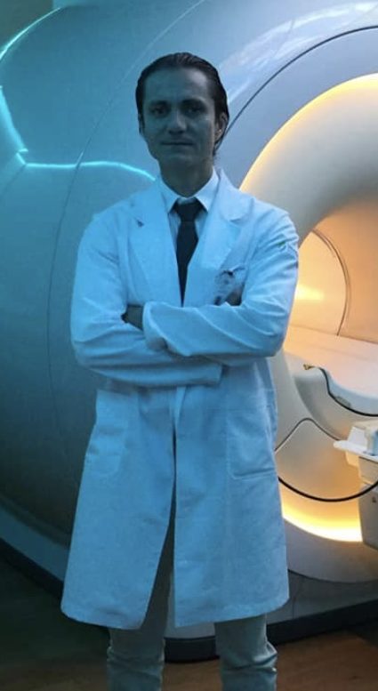 Prof. Juan Cosme is professor of magnetic resonance imaging at the National Institute of Nutrition Salvador Zubirán and associate professor of sports medicine, traumatology and minimally invasive sports surgery at the National Autonomous University of Mexico (Universidad Nacional Autónoma de México, UNAM) in Mexico City. He is part of the medical staff of the America Football Club and the Mexico National University Football Club, two local professional teams where he is in charge of diagnosing player’s injuries with imaging. Prof. Cosme performs special diagnostic studies with arthro-MRI and therapeutic procedures with infiltrations for specific musculoskeletal pathologies. He participates as a teacher in the courses given at the Orthopaedics and Radiology schools. He has published several book chapters on orthopaedics and sports medicine, and articles in radiological journals.