European Society of Radiology: Sports imaging is the main theme of IDoR 2019. In most countries, this is not a specialty in itself, but a focus within musculoskeletal radiology. In your country, is there a special focus on sports imaging within radiology training or special courses for interested radiologists?
Mamoru Niitsu: Recently, the number of Japanese musculoskeletal (MSK) radiologists has gradually increased; however, no training course that specifically focuses on sports imaging is provided at the majority of universities and hospitals. Of course, some training camps for young radiologists include short sports medicine courses that last a few days or weeks. MSK courses are still considered to be relatively minor courses among ‘major’ courses, including neurology, chest, cardiac, hepatobiliary, digestive tract, urogenital, and obstetrics and gynaecology courses.
ESR: Please describe your regular working environment (hospital, private practice). Does sports-related imaging take up all, most, or only part of your regular work schedule?
MN: I work at a university hospital with approximately 1,000 beds. I mainly engage in diagnostic MRI and CT imaging, covering the whole body from head to toe. I am responsible for reading all MSK images, which account for a fourth to a third of all images read in daily practice. Half of the MSK imaging performed in daily practice is sports-related imaging, for example, in patients with muscle strains, joint injuries, or traumatic bone injuries. The other half involves patients with osteoarthritis, rheumatoid arthritis, myopathy, diabetic foot injuries, etc.
ESR: Based on your experience, which sports produce the most injuries that require medical imaging? Have you seen any changes in this regard during your career? What areas/types of injuries provide the greatest challenge to radiologists?
MN: Contact sports, including rugby, football, and basketball, and events involving long periods of running, such as long-distance running, marathons, triathlons, and football, are particularly associated with leg injuries. With the recent marathon boom, many amateur runners complain of joint pain and muscle soreness. Football is the most popular sport among Japanese boys and girls, replacing baseball ten years ago. As a result, Osgood-Schlatter disease, Sinding-Larsen-Johansson disease, Jumper’s knee, and meniscal/ligament injuries now attract much attention. Recently, focal periphyseal oedema (FOPE) and Morel-Lavellee lesions have been included among the differential diagnoses for knee pain in young athletes. In addition, cartilage imaging of the hip, knee, and ankle have become more important.
ESR: Please give a detailed overview of the sports injuries with which you are most familiar and their respective modalities.
MN: I mainly diagnose articular cartilage damage. Current powerful MRI techniques can be used to perform internal evaluations, such as assessments of the glycosaminoglycan (GAG) concentration based on T2 mapping, T2* mapping, T1rho mapping, and Chemical Exchange Saturation Transfer (CEST). These qualitative imaging techniques are attracting great interest among radiologists, orthopaedic surgeons and pathologists.
ESR: What diseases associated with sporting activity can be detected with imaging? Can you provide examples?
MN: As mentioned above, cartilage damage can be detected with MRI. High-quality MRI images with sufficient spatial and contrast resolution can detect subtle cartilage surface irregularities. Moreover, qualitative imaging, such as T1rho mapping, can be used to evaluate internal changes in the cartilage layer, which makes it possible to assess on-going changes in the articular cartilage.
ESR: Radiologists are part of a team; for sports imaging this likely consists of surgeons, orthopaedists, cardiologists and/or neurologists. How would you define the role of the radiologist within this team, and how would you describe the cooperation between radiologists, surgeons, and other physicians?
MN: As they read images and make diagnoses, radiologists are often the first point of contact between patients and medical teams. Of course, all of the activity of medical teams is based on the initial diagnosis. Therefore, the role of radiologists is essential. In addition, radiologists act as coordinators of medical teams, directing further examinations and procedures.
ESR: The role of the radiologist in determining diagnoses with sports imaging is obvious; how much involvement is there regarding treatment and follow-up?
MN: Radiologists should monitor patients’ ongoing treatment and follow-up. Therefore, they should have frequent and close communication with surgeons and other members of staff.
ESR: Radiology is effective in identifying and treating sports-related injuries and diseases, but can it also be used to prevent them? Can the information provided by medical imaging be used to enhance the performance of athletes?
MN: Precise diagnosis and appropriate treatment are required to enhance the performance of athletes. As they initiate many of the activities of medical teams, radiologists, who have access to a variety of data, can direct the next steps in treating each patient as well as the future practice of medical teams. Of course, radiologists should provide careful physical and mental support to injured athletes.
ESR: Many elite sports centres use cutting-edge medical imaging equipment and attract talented radiologists to operate it. Are you involved with such centres? How can the knowledge acquired in this setting be used to benefit all patients?
MN: I work as a visiting radiologist at the Japan Institute of Sport Sciences (JISS), where training and medical check-ups of top athletes, including Olympic athletes, are performed. The Japanese government established the JISS in 2001. Since then, the Japanese team has achieved favourable results at the Olympic Games held in Athens, Beijing, London, Rio de Janeiro, and Pyeongchang. At the clinic within the JISS, cutting-edge medical imaging equipment, including two 3T MRI scanners, allows high-quality imaging. These sophisticated imaging techniques can be distributed to hospitals and clinics nationwide.
ESR: The demand for imaging studies has been rising steadily over the past decades, placing strain on healthcare budgets. Has the demand also increased in sports medicine? What can be done to better justify imaging requests and make the most of available resources?
MN: In Japan, all medical procedures are performed under the national health insurance system or personal health insurance coverage, except on rare occasions, for example when such procedures are performed for the JISS or a professional baseball/football team. As MRI and CT scanners are in very high demand, all imaging examinations have to be booked in advance. This can result in selection bias for such examinations, but it can also help to exclude unnecessary examinations. Radiologists should recommend the most effective and balanced examination pathway for each patient.
ESR: Athletes are more prone to injuries that require medical imaging. How much greater is their risk of developing diseases related to frequent exposure to radiation, and what can be done to limit the negative impacts from overexposure?
MN: In my field, MRI plays a significant role, and CT, a relatively small role, especially in joint and muscle imaging. Thus, the risk of excessive radiation exposure seems quite low.
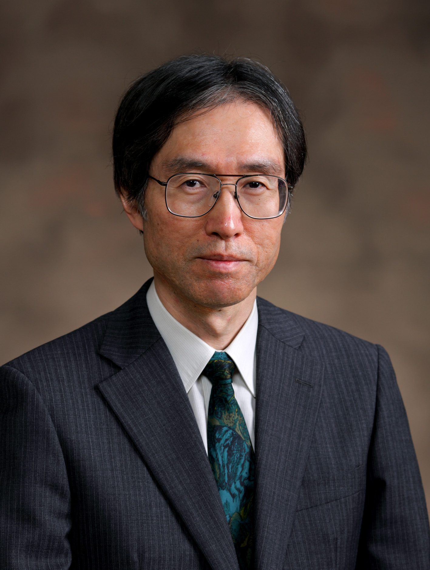
 Prof. Mamoru Niitsu is professor and chairman of the Department of Radiology, Saitama Medical University in Saitama, Japan. He graduated from Tsukuba University in 1986. After his residency training at the Department of Radiology, Tsukuba University Hospital, he joined the Magnetic Resonance Laboratory of the Department of Diagnostic Radiology at Mayo Clinic, USA, in 1991. He was involved in a number of MR research activities, such as kinematic joint imaging, muscle kinematic MRI and T1 calculation using tag-implementation. He continued and expanded MR research, especially in musculoskeletal imaging, after moving back to Tsukuba and Saitama. Recently, he worked to advance articular cartilage imaging using T1rho and CEST technique as well as ultrasonography of the joint imaging.
Prof. Niitsu was a president of the Japanese Society for Magnetic Resonance in Medicine (JSMRM) from 2012 to 2014, chair of the society’s annual meeting in 2016 and its international committee from 2016 to 2018. He sits on the editorial boards of European Radiology, Magnetic Resonance in Medical Sciences, and the Japanese Journal of Radiology.
He will serve as a chief musculoskeletal radiologist for the coming Tokyo 2020 Olympic/Paralympic Games.