European Society of Radiology: Sports imaging is the main theme of IDoR 2019. In most countries, this is not a specialty in itself, but a focus within musculoskeletal radiology. In your country, is there a special focus on sports imaging within radiology training or special courses for interested radiologists?
Norma Pedreañez: Currently, there are no accredited training or special courses on sports imaging available to radiologists in Venezuela. Three years ago, I designed a course in musculoskeletal imaging for the university I work at, to train postgraduate interns in radiodiagnostics, which included topics on sports imaging.
ESR: Please describe your regular working environment (hospital, private practice). Does sports-related imaging take up all, most, or only part of your regular work schedule?
NP: I work at two private centres (Grupo Óseo and Grupo Medimagen, both in Caracas) and both have image units. At Grupo Óseo, head-to-toe studies are carried out, but our strength there is the musculoskeletal system. In both institutions, I invest 80% of my time in musculoskeletal imaging, using x-ray, computed tomography (CT), ultrasound and magnetic resonance imaging (MRI). These two centres are reference centres in musculoskeletal ultrasound, arthro-MRI and arthro-CT.
ESR: Based on your experience, which sports produce the most injuries that require medical imaging? Have you seen any changes in this regard during your career? What areas/types of injuries provide the greatest challenge to radiologists?
NP: I have seen frequent injuries in patients practising baseball, football, tennis and running. Currently, I have seen more frequent injuries related to the disciplines of Total Resistance exercises (TRX) and cross-fit. I think that the greatest challenge for radiologists depends on their knowledge of the topic. Lesions of the deep subgluteus space, the ischio femoral impingement and the subspinosus clamping are traditionally not well known, and therefore underdiagnosed. The same is true for the interpretation of arthro-MRI and arthro-CT.
ESR: Please give a detailed overview of the sports injuries with which you are most familiar and their respective modalities.
NP: In baseball players, I most often see injuries of the glenoid labrum (SLAP), most frequently SLAP II and specifically SLAP IIB associated with internal posterior superior impingement. I also often diagnose SLAP IIA, IIC and IV; tendinitis of the biceps brachial; and intra-substance injuries of the biceps brachii in these patients.
To evaluate SLAP, the modality most used is arthro-MRI. In the case of suspected intra-articular injuries, we will carry out a simple MRI or arthro-MRI examination, and in the case of extra-articular injuries, an ultrasound scan.
In football players, I am familiar with traumatic lesions of the meniscus, such as meniscal contusion and ruptures, partial and complete ruptures of the ACL. For meniscus and cruciate lesions, we will perform a simple MRI examination. We also see quite a lot of proximal injuries of the tendons and adductor muscles and muscle tears, especially in quadriceps femoral and calf. In proximal injuries of the tendons and adductor muscles, we use ultrasound first, and in the case of suspected athletic pubalgia associated with that lesion, we do a simple MRI scan and ultrasound in case of muscle tears, quadriceps femoral, and gastrocnemius.
In tennis players, I am most familiar with muscle tears, triangular fibrocartilage injuries and ulnar extensor tendon of the carpus, and shoulder injuries, which are similar to baseball players.
In running injuries, I often have to deal with iliotibial band friction, Achilles ruptures, sesamoid fractures and tibial periostitis. In both cases, we first use ultrasound; for sesamoid fractures and tibial periostitis, a simple MRI scan.
ESR: What diseases associated with sporting activity can be detected with imaging? Can you provide examples?
NP: Imaging typically helps to detect injuries from chronic overuse that generally affect the lower limbs, obviously depending on the type of sport and discipline. To name a few: osteochondral lesions in the femoral condyles and the astragalus, tibial periostitis, athletic pubalgia (sports hernia); stress fractures in hallux sesamoid bones, calcaneus, plantar fasciitis; Achilles and patellar tendinopathies; and tendinous, ligamentary and acute muscular injuries.
ESR: Radiologists are part of a team; for sports imaging this likely consists of surgeons, orthopaedists, cardiologists and/or neurologists. How would you define the role of the radiologist within this team, and how would you describe the cooperation between radiologists, surgeons, and other physicians?
NP: Most pathologies in sports medicine often require an imaging test, so the role of the radiologist in diagnosing and monitoring these injuries is key. Multidisciplinary management is essential, and it is also important to understand lesion mechanism. It is necessary to be able to differentiate between findings of adaptive phenomena of the sport gesture, and to know the therapies and therapeutic procedures to be performed by the athlete or physiotherapist or in conjunction with other specialists, including radiologists, to be able to perform an adequate imaging analysis and follow-up.
ESR: The role of the radiologist in determining diagnoses with sports imaging is obvious; how much involvement is there regarding treatment and follow-up?
NP: Along with orthopaedic surgeons, we perform echo-guided punctures for bio supplementation therapies in muscle tears and intra-articular drainage collections. Athletes require a prompt and correct recovery; therefore the imaging follow-up complements the clinical follow-up and is key to maintain or make changes in physiotherapy, or highlight the need for surgical intervention, helping the athlete to return to practice.
ESR: Radiology is effective in identifying and treating sports-related injuries and diseases, but can it also be used to prevent them? Can the information provided by medical imaging be used to enhance the performance of athletes?
NP: Yes, the performance of athletes can be improved by taking into account the use of medical images. For example, we can evaluate the type of muscle fibre of an athlete (type 1, type 2), evaluate the trophism and tendons on ultrasound. Based on that information, we can contribute as radiologists in training planning.
ESR: The demand for imaging studies has been rising steadily over the past decades, placing strain on healthcare budgets. Has the demand also increased in sports medicine? What can be done to better justify imaging requests and make the most of available resources?
NP: Yes, I think there is an increase in the demand for imaging studies in sports medicine, which is due to the greater frequency of sports practice and the need to use imaging methods in therapeutics and follow-up. As radiologists, we must participate in the creation of study protocols, establishing the imaging modality that is suitable for each case.
ESR: Athletes are more prone to injuries that require medical imaging. How much greater is their risk of developing diseases related to frequent exposure to radiation, and what can be done to limit the negative impacts from overexposure?
NP: The suspected diagnosis determines the imaging technique to be used. Since soft tissue injuries are the most common, ultrasound is widely used, with the great advantage of being able to perform dynamic studies. MRI is very useful in the evaluation of intra-articular injuries, as well as bone lesions of stress. Both these modalities do not expose the athlete to ionising radiation.
ESR: Do you actively practise sports yourself and if yes, does this help you in your daily work as MSK radiologist?
NP: I do not practise any sports discipline in a ‘formal way’, but I practise trekking and regularly climb Avila Mountain. I go to the gym three times a week and particularly enjoy TRX and Pilates.
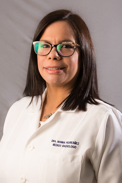 Dra. Norma Pedreañez is ad honorem Assistant Professor in the radiodiagnostics department of the Clinic University Hospital and ad honorem Professor in the shoulder and spine committees of the Children’s Orthopaedic Hospital and the radiodiagnostics graduate schools in Caracas, Venezuela. She works as a radiologist in a bone group where she specialises in musculoskeletal imaging. She trains orthopaedic traumatology fellows specialising in shoulder, hip and knee in musculoskeletal imaging. She also works at Grupo Óseo and Grupo Medimagen, two private centres in Caracas. Dr. Pedreañez undertook an extension course in tomography and magnetic resonance imaging at the Dr. Gilberto Rodriguez Ochoa Latin American Children’s Cardiology Hospital in Caracas. She received her diploma in magnetic resonance and Carema from the School of Engineering of Venezuela Central University. She is a regular speaker at national conferences and a member of the Venezuelan Society of Radiology and Radiodiagnostics and sits on the board of the Venezuelan Society of OrthoBiology.