European Society of Radiology: Could you please give a detailed overview of when and for which diseases you use cardiac imaging?
Adrian Santa: All diseases that benefit from cardiac imaging, starting with coronary CT (about six per day), looking for malformation of coronary origin and tract, stenosis, stent control, bypass control and transcatheter aortic valve implantation (TAVI) planning. The main field for cardiac imaging with MRI is in the field of cardiac malformations and follow-up after surgery, myocardial disease (dilative and hypertrophic cardiomyopathy, status after infarction-transmural or subendocardial, complications e.g. aneurysmal dilatation), myocarditis, amyloid, sarcoid, and post-surgery control. Also ruling out chest pain is a domain, taking into account that negative predictability of coronary CTA is very high, over 98%.
ESR: Which modalities are usually used for what?
AS: Coronary CTA is used to rule out chest pain, effort anger, malformations of origin and coronary tract, dissection, atheromatosis and stenosis of coronary, some malformations of the heart, also for stent and bypass control, TAVI planning, postoperative assessment (diameters of the pulmonary artery, aortic valve etc.). MRI is used to assess malformations, right ventricular arrhythmias, valvular pathology and mainly myocardial disease, e.g. myocarditis, infarction and its consequences, amyloidosis, sarcoid, cardiac tumours and cardiomyopathies.
ESR: What is the role of the radiologist within the ‘heart team’? How would you describe the cooperation between radiologists, cardiologists, and other physicians?
AS: The role of the radiologist is being ‘the eyes’ of the cardiologist, cardio-coronary imaging is prone to detect pathology, prepare for TAVI and surgery by giving anatomical data in malformations of heart and coronaries, so activity is in close conjunction with the cardiologist and cardiac surgeon both prior to and after surgery. We hold discussions on cases and perform reconstructions on graphic stations in order to establish planning and follow up.
ESR: Radiographers/radiological technologists are also part of the team. When and how do you interact with them?
AS: All the time. Technologists perform primary acquisition of images under close control from the radiologist and have a short discussion with the patient prior to the procedure to inform and instruct the patient about the examination, regarding movement, breathing and so on. The technologist sets the patient up (EKG, placement of venous catheter) and also tests for contrast intolerance.
ESR: Please describe your regular working environment (hospital, private practice). Does cardiac imaging take up all, most, or only part of your regular work schedule? How many radiologists are dedicated to cardiac imaging in your team?
AS: Apart from educational work at the university, my medical activity takes place in a private hospital. All kind of CT and MRI are performed, cardiological and vascular work being most prominent on CT, taking approx. 70% of CT. Cardiac MRI is done 2–3 times per week, at the clinician’s demand.
ESR: Do you have direct contact with patients and if yes, what is the nature of that contact?
AS: Prior to examination, each patient comes with the medical data that is studied by the radiologist, including allergies, values of creatinine and urea, psychical condition – some patients are scared of not knowing what the examination consists of. Everything is explained to the patient by the radiologist. At request, the results of the examination are discussed with the patient further after the exam.
ESR: If you had the means: what would you change in education, training and daily practice in cardiac imaging?
AS: Currently I am holding cardiac and coronary imaging courses for radiologists to increase the number of qualified radiologists in this field, and to reduce the turf war with cardiologists that try to take over cardiac imaging.
ESR: What are the most recent advances in cardiac imaging and what significance do they have for improving healthcare?
AS: Multislice fast CT with temporal and spatial resolution that allows consistent results in patients with up to 75 b/min of heart rate, and contrast media that is less allergenic (iso-osmolar substances with high iodine content). Graphic stations dedicated to cardiac and coronary reconstructions are available saving a lot of time. Fractional flow reserve (FFR) calculation with coronary CTA is a new field that hopefully will be available soon, offering haemodynamic data, very important in stenotic coronary disease.
ESR: In what ways has the specialty changed since you started? And where do you see the most important developments in the next ten years?
AS: Enormously, due to technical improvements of the industry and to a better understanding by clinicians of what we could provide for them.
ESR: Is artificial intelligence already having an impact on cardiac imaging and how do you see that developing in the future?
AS: Graphic stations that automatically do segmentation and labelling, stenosis quantification and FFR calculations are fields where advancements are welcome as they increase our capabilities of diagnosis and improve the likelihood of patients being examined daily by reducing the reconstruction times.
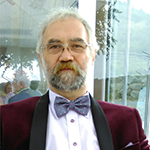
 Dr. Adrian Santa is Assistant Professor of Radiology at the Faculty of Medicine, ‘Lucian Blaga’ University in Sibiu, Romania, and he is also currently working as radiologist in a private hospital with emphasis on cardiology and cardiac surgery. A graduate of Medical University Tîrgu Mureş, his current practice includes CT and MRI in all domains. He specialised in radiology at Fundeni Clinical Hospital in Bucharest in 1995 and received his PhD in radiology and medical imaging in 2001 at the Medical University Cluj Napoca. Dr. Santa has held many courses in cardiac imaging mainly abroad (Vienna, Barcelona), and also holds courses twice a year for radiologists in CT coronarography and cardiac MRI. He is currently vice-president of the Romanian Society of Magnetic Resonance, member of the Board of the Romanian Society of Ultrasonography, member of the Romanian Society of Radiology and member of the European Society of Radiology, with many scientific presentations on cardiac imaging in congresses in Romania and abroad (the latest was at the Balkan Society of Radiology in Budapest in 2017 as an invited lecturer).