European Society of Radiology: Could you please give a detailed overview of when and for which diseases you use cardiac imaging?
Elena Mershina: Nowadays it is not possible to imagine the diagnosis and treatment of cardiac diseases without non-invasive cardiac imaging. Cardiac imaging is crucial for diagnosis and management of the patients with congenital heart disease, valvular pathology, ischaemic heart disease, myocardial and pericardial diseases (myocarditis and cardiomyopathies in particular). Patients with diseases of pulmonary arteries and aorta, cardiac and paracardiac masses also need cardiac imaging. It is used to establish and to estimate the severity of the disease, to define the treatment strategy and to observe the results of treatment.
ESR: Which modalities are usually used for what?
EM: Ultrasound (echocardiography) in clinical practice has been used as the first-line imaging modality in practically every case.
The main role of CT is the performance of non-invasive coronary angiography (CCTA). Modern guidelines say that in patients with a low or intermediate pre-test probability of obstructive CAD it is CCTA that can exclude or confirm the presence of obstructive coronary lesions. CCTA is recommended in patients with acute chest pain and low probability of acute coronary syndrome. CCTA is used as the ‘gold standard’ for non-invasive visualisation of the thoracic and abdominal aorta. It has become an accepted imaging standard for the planning of TAVI procedures.
Stress echocardiography, single-photon emission computed tomography (SPECT) and perfusion magnetic resonance imaging (MRI) are the cardiac imaging modalities traditionally used for non-invasive detection of myocardial ischaemia. They have already been included in different national and international cardiological and radiological guidelines.
Cardiac MR (CMR) is a method of choice for characterisation of the myocardium. It is used for diagnostic workup of cardiomyopathies, acute and chronic myocarditis, for detection and qualitative assessment of myocardial scars and fibrosis.
ESR: What is the role of the radiologist within the ‘heart team’? How would you describe the cooperation between radiologists, cardiologists, and other physicians?
EM: I believe that the radiologist plays a leading role in a cardiac team. A good cardiac radiologist can give the answers to all the cardiologist’s questions and provide crucial diagnostic information for the cardiac surgeon. But of course, he has to be a very good expert both in radiology and heart diseases.
ESR: Radiographers/radiological technologists are also part of the team. When and how do you interact with them?
EM: Radiographers play a very important role in providing the radiologist with the datasets of high-quality cardiac images. We work with them hand-to-hand every day. They prepare patients for the study, they perform CCTA and CMR under radiologist’s supervision and they care for patients after the end of the study. For the best results of teamwork, the radiologists should take care of permanent training and education of radiographers.
ESR: Please describe your regular working environment (hospital, private practice). Does cardiac imaging take up all, most, or only part of your regular work schedule? How many radiologists are dedicated to cardiac imaging in your team?
EM: I am Head of the Radiology Department in the University Hospital of Moscow State University. Cardiac imaging takes up a substantial part of my regular work schedule. In our country, we still do not have a formal division into subspecialties. So a cardiac radiologist is usually a general radiologist with special interest and training in one or several clinical areas. In our team, there are two more radiologists who can do cardiac imaging (CCTA and CMR) on the expert level. But most of our radiologists also can do basic cardiac studies.
ESR: Do you have direct contact with patients and if yes, what is the nature of that contact?
EM: Yes, we have direct contact with patients and I believe that it is a big advantage both for radiologists and patients. Usually, I speak with the patients personally before an examination in order to better understand the indications to the study, to look for the possible contra-indications. Sometimes after such an interview, I re-direct the patient to another type of cardiac examination which is more informative and appropriate for his case. After the examination, I explain in brief to the patient the preliminary results of the examination. Sometimes I even make special appointments with patients when there is a need for the detailed explanation of the radiological report, especially when there are some discrepancies between the results of different imaging tests. From time to time I also play the role of a cardiologist and give the patient some advice about options for further diagnostic steps and possible treatment.
ESR: If you had the means: what would you change in education, training and daily practice in cardiac imaging?
EM: I am not the first person who has talked about the necessity of longer training of residents in Russia. Today, the duration of residency for radiologists is two years and there is no formal subspecialty fellowships position after it. So I see a clear need for bringing our national residency training closer to the standards recommended by the ESR that are described in detail in the European Training Curriculum in Radiology. And of course, a programme of fellowship cardiac radiology must be created and launched. I and my colleague Professor Valentin Sinitsyn are going to develop and implement such a programme in the near future.
But in spite of all these drawbacks, anyone interested in cardiac imaging can learn a lot and get training by participation in different schools and courses, attending national and international congresses of radiology – and of course, ESR, ECR, ESOR, and ESCR activities are the most attractive and important ones for cardiac radiologists.
ESR: What are the most recent advances in cardiac imaging and what significance do they have for improving healthcare?
EM: Modern CT scanners have the opportunity to study rest and stress myocardial perfusion and to combine these data with the results of non-invasive CCTA. Such an approach improves the specificity of CCTA and decreases the need for cardiac catheterisations. There are also some other new technologies for the assessment of coronary blood flow, probably the most interesting one is a non-invasive assessment of fractional flow reserve by CT (FFR-CT) with the help of sophisticated computer analysis. The use of CCTA in clinical trials for the detection of vulnerable plaques and stratification of patients according to the severity of coronary atherosclerotic burden has already brought very promising results. Dual-energy CCTA could be used for myocardial characterisation practically for the same indications as CMR – e.g. for the detection of post-infarction myocardial scars, myocarditis, and cardiac amyloidosis. Besides this, dual-energy coronary CT helps to eliminate artefacts from calcified plaques obscuring the lumen of coronaries, metallic stents, and other implants.
Besides coronary and perfusion imaging, CMR (a recognised reference standard for myocardial imaging) is a standard tool for the assessment of non-coronary myocardial diseases. T1- and T2-mapping attracts a lot of attention as a unique modality to detect and quantify diffuse myocardial fibrosis.
Today it has become obvious that cardiac imaging may have an impact on a patient’s prognosis and the selection of the optimal treatment plans.
ESR: In what ways has the specialty changed since you started? And where do you see the most important developments in the next ten years?
EM: I started to do CMR and CCTA in the mid-90s. Looking back, I clearly see that cardiac imaging has changed dramatically. Recent changes in cardiological paradigms about approaches to diagnosis, treatment, and assessment of prognosis in patients with coronary artery disease (CAD ) together with the technical development of CCTA and accumulation of scientific data proving the high diagnostic value of CCTA have resulted in very interesting perspectives concerning the use of this modality for non-invasive coronary imaging. I believe that in the next ten years, myocardial CT-perfusion will become a routine procedure. The role of CMR in arrhythmology is becoming more and more visible. It is used before radiofrequency ablation to improve its safety and to exclude non-necessary procedures.
ESR: Is artificial intelligence already having an impact on cardiac imaging and how do you see that developing in the future?
EM: It depends what you mean using the term artificial intelligence (AI). For a long time, a cardiac workstation has been capable to automatically detect heart chambers and vessels, label and segment them, automatically calculate volumes and other quantitative parameters. Today we take such tools for everyday practice for granted. Now we are speaking about AI as a reliable tool for the performance of more complicated tasks including assessment of diagnosis and prognosis. In this sense, there is no big impact of AI on cardiac imaging right now. But AI is developing and I see our task in finding the most useful areas in its application to cardiac imaging.
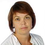
 Dr. Elena Mershina is Associate Professor of Radiology and Head of the Radiology Department at Moscow State University in Moscow, Russia. She is an ESR member, a board member of the ESCR, the Russian Society of Radiology and the Russian Society of Cardiovascular Radiology. She is an internationally renowned expert in cardiac imaging. Her main research interest is CMR in cardiomyopathies, searching of radiologic-genetic correlations; CMR and CCTA in CAD; MRI and dual-energy CT (DECT) in pulmonary hypertension.
Dr. Mershina has authored and co-authored 28 peer-reviewed publications and has given numerous invited lectures at national and international meetings (ECR 2013, 2014, ESCR 2016, 2017, ESOR 2018). During ECR 2018 she organised and moderated the multidisciplinary session ‘The heart team: coronary imaging and treatment’.
From 2011 to 2013 she was an ECR cardiac subcommittee member, from 2014 to 2016 thoracic subcommittee member.