European Society of Radiology: Could you please give a detailed overview of when and for which diseases you use cardiac imaging?
Hatem Alkadhi:
- Stable coronary artery disease (CAD): ruling-out CAD, in- and out-patients
- Ruling-out CAD in patients prior to non-coronary cardiac surgery
- Assessment of aortocoronary bypass grafts
- Assessment of the anatomy of the left atrium and ruling-out left atrial thrombus
- Acute chest pain: triple rule-out CT or cardiac CT, patients from the emergency department or from the intensive care unit (chest pain unit)
- Grown-up congenital heart disease: in- and out-patients
- Coronary anomalies: in- and out-patients
- Assessment of myocardial ischaemia and infarction: in- and out-patients
- Peri/Myocarditis and storage diseases: mainly in-patients
- Cardiac tumours: both in- and out-patients
- Imaging prior to and after transcatheter aortic valve implantation (TAVI)
- Cardiomyopathy: both in- and out-patients
ESR: Which modalities are usually used for what?
HA: We use cardiac CT and cardiac MR imaging. Echocardiography and catheter coronary angiography is done by cardiologists only.
We use the two modalities as follows:
- Cardiac CT and cardiac MRI: Stable coronary artery disease (CAD): ruling-out CAD
- Cardiac CT: ruling-out CAD in patients prior to non-coronary cardiac surgery
- Cardiac CT: assessment of aortocoronary bypass grafts
- Cardiac CT: assessment of the anatomy of the left atrium and ruling-out left atrial thrombus
- Cardiac CT: acute chest pain: triple rule-out CT or cardiac CT, patients from the emergency department or from the intensive care unit (chest pain unit)
- Cardiac CT and cardiac MRI: grown-up congenital heart disease
- Cardiac CT: coronary anomalies
- Cardiac MRI: assessment of myocardial ischaemia and infarction
- Cardiac MRI: peri/myocarditis and storage diseases
- Cardiac MRI and CT: cardiac tumours
- CT: Imaging prior to and after transcatheter aortic valve implantation (TAVI)
- Cardiac MRI: cardiomyopathy
ESR: What is the role of the radiologist within the ‘heart team’? How would you describe the cooperation between radiologists, cardiologists, and other physicians?
HA: We (the radiologists) perform and read all cardiac CT studies. We perform and read all cardiac MR imaging studies together with a cardiologist, who has a 50% appointment in our radiology department.
Collaboration and interaction with cardiologists and cardiac surgeons is crucial (as is in all other subdisciplines of radiology) for achieving and maintaining high standards of quality, and for understanding the demands of the clinicians.
ESR: Radiographers/radiological technologists are also part of the team. When and how do you interact with them?
HA: Radiographers/radiological technologists play perhaps the most important part in the entire chain from instructing and scanning patients until image interpretation, because only with well-trained radiographers/radiological technologists will we acquire good images. Without an adequate quality of the CT and MR images, no radiologist, whatsoever their skills are, can perform a good report and provide a correct differential diagnosis. Therefore, collaboration and extensive exchange between radiographers/radiological technologists and radiologists is the mainstay for running a successful imaging department. This is perhaps even more important in cardiac imaging than in other radiological subdisciplines because cardiac imaging is also technically quite demanding (maximal temporal and spatial resolution, ECG-gating, medication such as adenosine, dobutamine, nitro-glycerine …).
ESR: Please describe your regular working environment (hospital, private practice). Does cardiac imaging take up all, most, or only part of your regular work schedule? How many radiologists are dedicated to cardiac imaging in your team?
HA: I work in a University Hospital. Cardiac imaging takes a part of my daily clinical duties and emergency work. We have a core team of three seniors and five juniors who do the regular work. In the emergency setting each radiologist reads cardiac CT and triple-rule-out CT studies.
ESR: Do you have direct contact with patients and if yes, what is the nature of that contact?
HA: We speak with each patient who comes for a cardiac CT imaging study, in order to understand their complaints and the indication for CT, and – most importantly – to instruct them about the subsequent examination (breath-hold commands, heating sensations from contrast media injection, nitro-glycerine application). Similar to cardiac CT, there is close communication with the patient for briefing them regarding the upcoming examination (medication, breath-hold commands, effects from adenosine etc.) in cardiac MR.
ESR: If you had the means: what would you change in education, training and daily practice in cardiac imaging?
HA: I would ask all my radiological colleagues to seek a close collaboration with their clinical colleagues (cardiologists, cardiac surgeons), and to intensify the contact to the patients (so that the patients realise what the role of a radiologist is and that he is a doctor as well).
Regarding training and practice: I would not change too many things, structures and machines are quite good, it is all about fostering the interests of our young colleagues in cardiac imaging, to motivate our residents in gaining subspecialty expertise, and ultimately to perform the European Board of Cardiac Radiology (EBCR) diploma of the ESR.
ESR: What are the most recent advances in cardiac imaging and what significance do they have for improving healthcare?
HA: Cardiac CT today is moving from an anatomical modality towards a functional imaging modality, where we cannot assess only the morphology of coronary arteries (for determining stenosis of vessels) but also the effect of stenoses on coronary flow and myocardial perfusion. Coronary flow can be assessed via computational fluid dynamics (CFD) calculations for determining the fractional flow reserve (FFR) in a non-invasive manner. With this, we cannot only determine the degree of stenosis but also its effect on coronary flow, which indicates whether or not a stenosis is truly haemodynamically significant. The other option, myocardial perfusion imaging, recently became feasible with CT. Here, CT myocardial perfusion imaging offers the possibility to directly visualise and quantify the extent and degree of myocardial perfusion deficits resulting from coronary stenoses. Both CT FFR and CT myocardial perfusion imaging are somewhat competitive with each other, and the near future will show which one will finally enter daily clinical routine.
Cardiac MR imaging today represents a validated, non-invasive diagnostic imaging tool playing a major role in the assessment of cardiac morphology and function, including tissue characterisation. Current trends in cardiac MR imaging are T1 and T2 mapping techniques which offer a quantitative assessment of the myocardium. This may be helpful in evaluating both focal and diffuse myocardial disease such as fibrosis and oedema. These techniques have the potential to represent clinical imaging markers for long-term follow-up of patients with myocardial disease.
ESR: In what ways has the specialty changed since you started? And where do you see the most important developments in the next ten years?
HA: Compared to the early days where cardiac imaging was a cutting-edge technique for dedicated patients and experts in image interpretation, today cardiac imaging with CT and MRI has become robust, accurate and precise and hence can be applied to many patients even in an emergency setting, which has become a clinical routine. Recent advances in both cardiac CT and MRI show that the two modalities will prosper further and will serve as imaging biomarkers in a multitude of different diseases, providing both morphological and functional information at (regarding CT) very low radiation doses or (regarding MRI) absent burden to our patients.
ESR: Is artificial intelligence already having an impact on cardiac imaging and how do you see that developing in the future?
HA: I believe and hope that artificial intelligence will have an impact on cardiac imaging, mainly through automatisation of certain tasks, such as e.g. the automated segmentation of ventricles. Also, computer-aided detection of disease may be facilitated via artificial intelligence-driven software solutions. I look forward to these things to happen, as we are faced with increasing numbers of examinations and images almost every day, which we cannot manage anymore with sufficient speed and quality without the assistance from computers. We have a highly active research group dealing with this issue which has published already several scientific articles regarding the potential of these promising algorithms (see e.g. Mannil et al. Invest Radiol 2018;53:338-343 or Baessler et al. Radiology 2018;286:103-112).
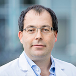
 Professor Dr. Hatem Alkadhi is a cardiovascular and emergency radiologist at the University Hospital Zurich, Switzerland. He trained in Zurich in general radiology and neuroradiology and pursued a fellowship in Boston (Massachusetts General Hospital) in cardiac radiology. He has a Master in Public Health from the Harvard School of Public Health (Boston, USA). Since 2002 he has been an active researcher in multimodality cardiovascular radiology, with a special focus on CT. Under his supervision, the Institute of Diagnostic and Interventional Radiology was the first worldwide to introduce cardiac CT into clinical routine.
Professor Alkadhi is an active speaker in Europe, Asia and the USA, where he has been invited to give more than 150 presentations and workshops. He is the author of 392 scientific papers, more than 30 book chapters and is editor of five books. He is the current President of the Swiss Society of Radiology (SGR-SSR).