European Society of Radiology: Could you please give a detailed overview of when and for which diseases you use cardiac imaging?
José Briceño Polacre: In our practice, the most common use is screening, especially in patients with high or intermediate risk of adverse cardiovascular events. Another important indication is to rule out coronary artery disease (CAD) that includes stenosis; those are the patients who regularly visit the doctor or the emergency room with atypical symptoms, unclear stress test results or electrocardiographic changes, chest pain with intermediate pre-test probability of CAD, evaluation of coronary stents and bypass grafts, suspicion of coronary anomalies, cardiac masses, pericardial disease, anatomy assessment in congenital heart disease, cardiomyopathy and electrophysiology.
ESR: Which modalities are usually used for what purpose?
JBP: For all the scenarios mentioned above we use cardiac computed tomography (CCT). In the case of calcium scoring, we use it to measure the presence of coronary calcifications, becoming a predictor of cardiovascular events. This technique does not require the use of intravenous contrast media.
ESR: What is the role of the radiologist within the ‘heart team’? How would you describe the cooperation between radiologists, cardiologists, and other physicians?
JBP: When we started doing CCT in 2009, cooperation was a bit tough, especially for some techniques like virtual colonoscopy, which also involves gastroenterologists. In most imaging centres in Venezuela, the radiologist took the leading role in CCT and cardiac magnetic resonance (CMR). In the beginning, we received more patients who had been referred by general practitioners, family doctors, internal medicine physicians and endocrinologists. After a year, we proved the validity of these methods, which produced interest from cardiologists and even interventional cardiologists and they started to get involved too. Today, in most centres, they play an equal role to the radiologists’.
ESR: Radiographers/radiological technologists are also part of the team. When and how do you interact with them?
JBP: Yes, radiographers and radiological technologists are a very important part of the team. They prepare the patient the day of the study, to have everything ready for scanning. The radiologist is present with the radiographer from the moment of acquiring the data and injecting contrast media. In some cases, once the raw data is obtained, the reconstruction of the final images is done with radiographers. But in my case, I reconstruct studies myself.
ESR: Please describe your regular working environment (hospital, private practice). Does cardiac imaging take up all, most, or only part of your regular work schedule? How many radiologists are dedicated to cardiac imaging in your team?
JBP: I work in a university hospital with a medical school and postgraduate programme, and a private practice. Cardiac imaging is only a part of what we do in daily practice. In the team, we are nine radiologists, three of whom do cardiac imaging.
ESR: Do you have direct contact with patients and if yes, what is the nature of that contact?
JBP: Yes we have direct contact with the patient. Before the procedure, we carry out an interview to highlight their cardiac background, and to explain the risks and benefits of what we are going to do, and what the procedure is going to feel like. On the day of the study, we are always present with the radiographer.
ESR: If you had the means: what would you change in education, training and daily practice in cardiac imaging?
JBP: I would include more cardiac anatomy and stress the correlation with medical imaging in the last years of medical school. We should also teach and encourage the use of low dose protocols at all times among the radiographers and radiologists, and promote the use of cardiac imaging as a method of screening among the population.
ESR: What are the most recent advances in cardiac imaging and what significance do they have for improving healthcare?
JBP: In the field of CCT, the most relevant advances are: faster scanners, motion correction algorithms, CT perfusion, metal artefact reduction software, structural heart planning software, iterative reconstruction and some other tools that allow us to do studies with high spatial and temporal resolution and a low dose of radiation. I consider CCT to be the most rapidly developing imaging technique in the diagnosis of CAD. It helps improve healthcare as a less invasive, reliable method with a high diagnostic value, and provides prognostic information for prediction of adverse cardiac events.
ESR: In what ways has the specialty changed since you started? And where do you see the most important developments in the next ten years?
JBP: In my opinion, the game changer is the ability to carry out CCT with low radiation dose and high diagnostic quality. Another thing that has changed is that cardiac imaging is popular among radiologists, who show more interest in all the cardiac imaging modalities, especially CCT and CMR. For the general practice of CCT, we use machines that are built for general purpose, but the immediate future is cardiac specific computed tomography machines. I also think that some other contrast materials will show up as an alternative to iodinated contrast. In some other modalities like MR, we will see better coils and faster machines that might not need contrast media at all or the incorporation of biomarkers in routine scans.
ESR: Is artificial intelligence already having an impact on cardiac imaging and how do you see that developing in the future?
JBP: 2017 will be remembered as the year in which artificial intelligence (AI) exploded in radiology. AI is here to support and increase the work of radiologists and not to replace them. AI will impact all human areas, including healthcare and of course cardiac imaging. We have seen in recent years how several automated methods have been developed to standardise the evaluation of calcified and non-calcified plaque and the lumen of the vessel. Several algorithms to evaluate coronary calcium semi-automatically and automatically have been proposed. Other automations include the ones that can estimate fractional flow reserve (FFR) by computational processing from coronary CTAngio. Other uses that are being evaluated for semi-automated measurement are perfusion and quantification of coronary epicardial fat. In this sense, radiologists cannot look away and say AI will not work; it is appropriate to adopt this technology and use it for the benefit of the specialty and in particular cardiac imaging.
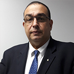
 Professor Jose Briceño Polacre is the chief medical radiologist at the Valera University Hospital in Venezuela. He is a professor of radiology for undergraduate and graduate students at the University of the Andes. He is also a professor at the Colegio Interamericano de Radiología (CIR).
Professor Briceño Polacre received general radiology training at the University Hospital of Maracaibo, attached to the University of Zulia, where he also obtained his PhD in medical science. He trained in vascular Doppler at the San Carlos Clinical Hospital in Madrid and did a fellowship in cardiovascular computed tomography (CCT) at Atlantic Medical Imaging New Jersey, US.
Professor Briceño Polacre obtained Level III Certification and received fellowship of the Society of Cardiovascular Computed Tomography (SCCT) in 2011 for his outstanding contribution in cardiovascular disease through CCT research, education and patient care.
He is an active speaker in Venezuela and Latin America and has more than 133 publications, scientific posters and conferences. His major interest is to promote the use of low dose of radiation in computed tomography, especially in cardiac imaging.
He is currently a corresponding member of the National Academy of Medicine of Venezuela, Vice President of the Bolivarian Region of CIR, Member of the Executive Committee of LatinSafe and President of the Venezuelan Society of Radiology and Diagnostic Imaging (SOVERADI).