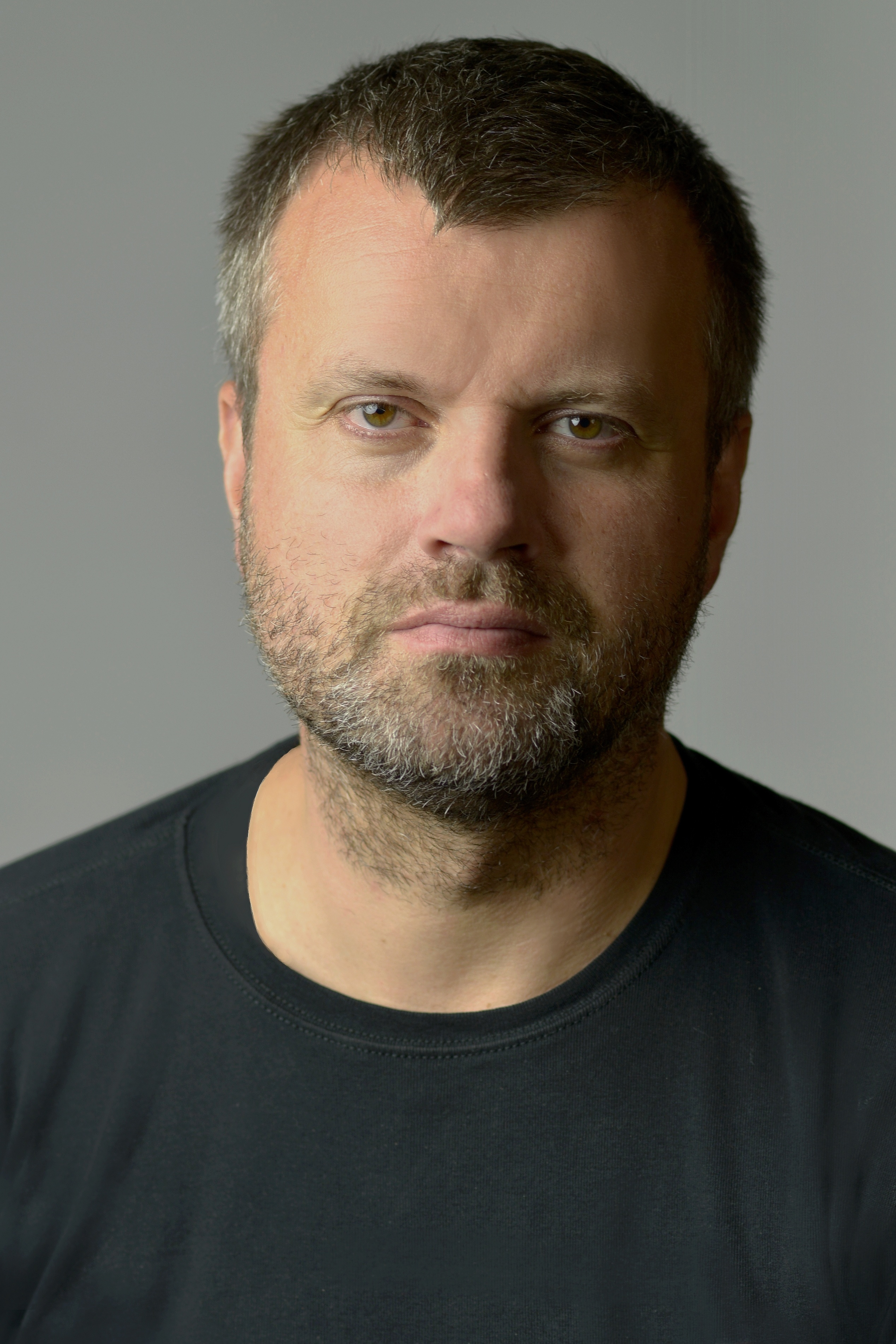
European Society of Radiology: Sports imaging is the main theme of IDoR 2019. In most countries, this is not a specialty in itself, but a focus within musculoskeletal radiology. In your country, is there a special focus on sports imaging within radiology training or special courses for interested radiologists?
Vladimír Neuschl: Musculoskeletal (MSK) radiology and sports imaging as its sub-discipline are included in the overall postgraduate radiology training in Slovakia. Several thematic and interdisciplinary professional events are organised in this area. Recently, a separate section of MSK radiology has been established in the Slovak Radiological Society.
ESR: Please describe your regular working environment (hospital, private practice). Does sports-related imaging take up all, most, or only part of your regular work schedule?
VN: I work at two private radiological centres where MSK imaging is a part of my daily routine. We try to carry out the examinations related to sports injuries as high priority cases. Sports practitioners and other professionals mainly request magnetic resonance (MR) and ultrasound (US) exams. We approach each case individually, modify the exam protocols and add other imaging modalities. Joint and muscular exams comprise approximately 25% of all cases. The other type of service we offer is a second look at the provided documentation of examined athletes, where in many cases the outside interpretation does not correlate with their clinical findings.
ESR: Based on your experience, which sports produce the most injuries that require medical imaging? Have you seen any changes in this regard during your career? What areas/types of injuries provide the greatest challenge to radiologists?
VN: Football is a popular sport throughout the year in Slovakia; ice hockey and skiing in winter. These sports bring us the most examined patients with the most common indication being an injured knee joint. In the past, the most typical diagnosis of athletes was the unhappy triad of the knee, where ACL mono trauma is mainly detected. I consider ankle trauma evaluation and accompanying impingement syndromes to be one of more challenging, complicated diagnostics. These, combined with multi-modal imaging, are often required here.
ESR: Please give a detailed overview of the sports injuries with which you are most familiar and their respective modalities.
VN: As mentioned previously, the most common trauma indicating the use of MR are acute knee injuries in football with meniscoligamentous lesions. The shoulder is affected in contusion hockey injuries with AC joint lesions. Superior labral tear from anterior to posterior (SLAP) lesions in basketball and volleyball are prevalent, often requiring MR diagnostics. An anterior tibiofibular (ATFL) rupture of the ankle and calf with gastrocnemius medial head muscle strain are universal multi-sport injuries. Calf diagnostics and findings controls are carried out by ultrasound.
ESR: What diseases associated with sporting activity can be detected with imaging? Can you provide examples?
VN: Stress fractures have become increasingly interesting, especially of the ribs, since they are often not detected by an x-ray screening. In recent years, we have examined several recreational and professional top athletes, e.g. tennis players, or other athletes. These have mostly been women suffering from overtraining at the gym. They are often sent to us with an indication of chest and dorsal back pain. By consulting the patient and the modifying MR protocols, we have detected stress fractures several times. It is necessary to rule out ‘the female athlete triad,’ those being eating disorders, amenorrhea and osteoporosis.
ESR: Radiologists are part of a team; for sports imaging this likely consists of surgeons, orthopaedists, cardiologists and/or neurologists. How would you define the role of the radiologist within this team and how would you describe the cooperation between radiologists, surgeons, and other physicians?
VN: While working on my PhD, I was fortunate enough to collaborate with cardiologists when determining the arrhythmogenic substrate in the hearts of at risk patients using cardiac MR. Abnormal ECG patterns are present in 40% of athletic people; therefore, our task is to define a possible arrhythmogenic substrate with prognostic significance in the heart by using CT/MR cardiac examinations. This arrhythmogenic substrate results in left ventricle remodelling and hypertrophy as well as pathological functional parameters of the right ventricle with respect to arrhythmogenic right ventricular dysplasia (ARVD) detection. The detection of fibrosis in the left ventricle is a distinctive specification. As the only centre in Slovakia, we are able to quantify the amount of fibrosis in the heart by using late gadolinium enhancement (LGE) imaging in high-risk patients such as differential diagnosis of athletic heart and hypertrophic cardiomyopathy in athletes. In recent years, we have had very good experience in cooperation with dynamically developing physical therapy centres in Slovakia. For physical therapists, our diagnostic view of sports injuries is often necessary in their treatment.
ESR: The role of the radiologist in determining diagnoses with sports imaging is obvious; how much involvement is there regarding treatment and follow-up?
VN: We must carry out early, quality diagnostic imaging to identify athletes’ injuries, thus expediting the treatment process. At the present time, patients are becoming more demanding and shared decision making is a mainstay in the management of elite sports. MSK radiologists who deal with an injured athlete need to be a part of the medical team and need to feel a sense of responsibility. The key to success is a broad knowledge of the pathophysiology of acute sports injuries as well as chronic overuse injuries. A personal interview with a patient is crucial not only for establishing the primary radiological diagnosis, but at follow-up appointments as well.
ESR: Radiology is effective in identifying and treating sports-related injuries and diseases, but can it also be used to prevent them? Can the information provided by medical imaging be used to enhance the performance of athletes?
VN: I have already referred to the prognostic and preventative significance of cardiovascular CT/MR diagnostics in athletes where in some cases, our pathological findings indicate a high risk of SCD. Naturally, assessing subtle congenital MSK abnormalities and older injuries with our technology also helps to determine potential athletic performance levels.
ESR: Many elite sports centres use cutting-edge medical imaging equipment and attract talented radiologists to operate it. Are you involved with such centres? How can the knowledge acquired in this setting be used to benefit all patients?
VN: In the past, I worked at a specialised orthopaedic sports clinic where radiological diagnostics was based on low field joint MRI technology and x-ray, which made the MSK diagnostics very indicative. At present, in our radiological centres, we have the most modern 1.5 and 3T MR equipment with a switch indication option, e.g. for wrist imaging I prefer a 3T MR device with a dedicated wrist coil. Even the ultrasound machines are equipped with the most modern linear probes.
ESR: The demand for imaging studies has been rising steadily over the past decades, placing strain on healthcare budgets. Has the demand also increased in sports medicine? What can be done to better justify imaging requests and make the most of available resources?
VN: Issues with the financing of MSK examinations have been occurring over the last five years after a reduction in health insurance payments. In some radiological centres, the examinations are artificially re-scheduled in favour of others, e.g. neuroradiological exams. Fortunately, we were prepared for these changes by using quality diagnostic modalities. We have fairly new MR devices with which we are able to set the exam protocols, e.g. joints in under ten minutes, while maintaining excellent image quality. Also, our well-trained medical professionals are able to accelerate the pre and post-processing of MSK diagnostics.
ESR: Athletes are more prone to injuries that require medical imaging. How much greater is their risk of developing diseases related to frequent exposure to radiation and what can be done to limit the negative impacts from overexposure?
VN: I do not believe there are any risks associated with radiation exposure to athletes. There are other groups of patients e.g. with pulmonary disease where CT screenings are much more frequent. We rarely supplement MR or ultrasound athletic diagnostics with a CT examination with a 3D reconstruction for the clinicians. Unsatisfactory, repeated x-ray examinations, e.g. at the suspected stress fracture, are contraindicated and MR imaging is recommended instead.
ESR: Do you actively practise sports yourself and if yes, does this help you in your daily work as MSK radiologist?
VN: Alas, my interest in sports is purely theoretical.
European Society of Radiology: Sports imaging also applies to sports-related injuries of the brain. In case you are familiar with this, please also answer the following questions:
ESR: Which sports have the highest risk of inducing brain injuries?
VN: We encounter them in kickboxing and traditional boxing.
ESR: What imaging modalities do you use with traumatic brain injury specifically in athletes?
VN: The most frequently used modality is a brain MRI examination with an expanded traumatic protocol without administering a contrast agent.
ESR: What can be learned from sports-related injuries that can be applied to a broader use, for example those sustained through automobile or other accidents that cause traumatic brain injury?
VN: In our centres, we have limited experience in dealing with repeated post traumatic brain changes sustained in sport. Rather, we monitor patients after singular post traumatic brain injuries, e.g. car accidents. Interesting findings are in cases of CTE (chronic traumatic encephalopathy) in athletes. These are athletes that have had a history of repetitive brain trauma. More importantly, a history of repetitive brain trauma may also lead to other neurodegenerative disorders such as Parkinson’s disease, frontotemporal lobar degeneration, as well as other neurodegenerative diseases. These findings help us to refine the aetiology of the emergence and treatment of these diseases.
ESR: How have advances in brain imaging allowed you to predict patient outcomes more accurately?
VN: Of great prognostic significance is the detection of microhaemorrhages in the brain, which are detected by the susceptibility-weighted imaging (SWI) sequence. It turns out that the number and volume of microhaemorrhages identified using SWI have been shown to be correlated with neuropsychological and clinical outcomes.
 Dr. Vladimír Neuschl, MD, PhD is a general radiologist, Head of the Institute of Diagnostic Imaging in Trnava. He graduated from the Faculty of Medicine in Bratislava. Since 1999, he has been an active researcher in multimodality cardiac and musculoskeletal radiology with special interest in MRI and rich lecture activity at domestic and foreign seminars. He has published work within interdisciplinary research alongside his PhD study of the issue of cardiac imaging by magnetic resonance and the use of MR imaging in traumatology. He holds the position of a Chairman of the Cardiovascular Radiology Section of the Slovak Cardiac Society. He is a co-founder of the Slovak MSK section of the Slovak Radiology Society and a member of the European Society of Radiology (ESR) and the European Society of Cardiovascular Radiology (ESCR).
He is notable for his professional participation in founding one of the first MR centres in Slovakia, as well as having had the opportunity to be present at the inception of MR cardiovascular and musculoskeletal diagnostics in Slovakia.